3X0Q | pdb_00003x0q

ADP ribose pyrophosphatase from Thermus thermophilus HB8 in ESMM-state at reaction time of 20 min


Experimental Data Snapshot

  • Method: X-RAY DIFFRACTION
  • Resolution: 1.14 Å
  • R-Value Work: 
    0.147 (DCC) 
  • R-Value Observed: 
    0.153 (Depositor) 

Starting Model: experimental
View more details

wwPDB Validation 3D Report Full Report

Validation slider image for 3X0Q

Ligand Structure Quality Assessment 


This is version 1.3 of the entry. See complete history

Literature

ADP-Ribose Pyrophosphatase Reaction in Crystalline State Conducted by Consecutive Binding of Two Manganese(II) Ions as Cofactors

Furuike, Y.Akita, Y.Miyahara, I.Kamiya, N.

(2016) Biochemistry 55: 1801-1812

  • DOI: https://doi.org/10.1021/acs.biochem.5b00886
  • Primary Citation Related Structures: 
    3X0I, 3X0J, 3X0K, 3X0L, 3X0M, 3X0N, 3X0O, 3X0P, 3X0Q, 3X0R, 3X0S

  • PubMed Abstract: 

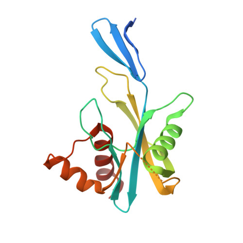
    Adenosine diphosphate ribose pyrophosphatase (ADPRase), a member of the Nudix family proteins, catalyzes the metal-induced and concerted general acid-base hydrolysis of ADP ribose (ADPR) into AMP and ribose-5'-phosphate (R5P). The ADPR-hydrolysis reaction of ADPRase from Thermus thermophilus HB8 (TtADPRase) requires divalent metal cations such as Mn(2+), Zn(2+), or Mg(2+) as cofactors. Here, we report the reaction pathway observed in the catalytic center of TtADPRase, based on cryo-trapping X-ray crystallography at atomic resolutions around 1.0 Å using Mn(2+) as the reaction trigger, which was soaked into TtADPRase-ADPR binary complex crystals. Integrating 11 structures along the reaction timeline, five reaction states of TtADPRase were assigned, which were ADPRase alone (E), the ADPRase-ADPR binary complex (ES), two ADPRase-ADPR-Mn(2+) reaction intermediates (ESM, ESMM), and the postreaction state (E'). Two Mn(2+) ions were inserted consecutively into the catalytic center of the ES-state and ligated by Glu86 and Glu82, which are highly conserved among the Nudix family, in the ESM- and ESMM-states. The ADPR-hydrolysis reaction was characterized by electrostatic, proximity, and orientation effects, and by preferential binding for the transition state. A new reaction mechanism is proposed, which differs from previous ones suggested from structure analyses with nonhydrolyzable substrate analogues or point-mutated ADPRases.


  • Organizational Affiliation
    • Graduate School of Science, Osaka City University , 3-3-138 Sugimoto, Sumiyoshi, Osaka 558-8585, Japan.

Macromolecule Content 

  • Total Structure Weight: 20.2 kDa 
  • Atom Count: 1,490 
  • Modeled Residue Count: 154 
  • Deposited Residue Count: 170 
  • Unique protein chains: 1

Macromolecules

Find similar proteins by:|  3D Structure
Entity ID: 1
MoleculeChains  Sequence LengthOrganismDetailsImage
ADP-ribose pyrophosphatase170Thermus thermophilus HB8Mutation(s): 0 
Gene Names: TTHA0528
EC: 3.6.1.13
UniProt
Find proteins for Q5SKW5 (Thermus thermophilus (strain ATCC 27634 / DSM 579 / HB8))
Explore Q5SKW5 
Go to UniProtKB:  Q5SKW5
Entity Groups
Sequence Clusters30% Identity50% Identity70% Identity90% Identity95% Identity100% Identity
UniProt GroupQ5SKW5
Sequence Annotations
Expand
Reference Sequence

Small Molecules

Ligands 4 Unique
IDChains Name / Formula / InChI Key2D Diagram3D Interactions
AR6

Query on AR6


Download:Ideal Coordinates CCD File
B [auth A][(2R,3S,4R,5R)-5-(6-AMINOPURIN-9-YL)-3,4-DIHYDROXY-OXOLAN-2-YL]METHYL[HYDROXY-[[(2R,3S,4R,5S)-3,4,5-TRIHYDROXYOXOLAN-2-YL]METHOXY]PHOSPHORYL] HYDROGEN PHOSPHATE
C15 H23 N5 O14 P2
SRNWOUGRCWSEMX-ZQSHOCFMSA-N
SO4

Query on SO4


Download:Ideal Coordinates CCD File
G [auth A]SULFATE ION
O4 S
QAOWNCQODCNURD-UHFFFAOYSA-L
GOL

Query on GOL


Download:Ideal Coordinates CCD File
F [auth A]GLYCEROL
C3 H8 O3
PEDCQBHIVMGVHV-UHFFFAOYSA-N
MN

Query on MN


Download:Ideal Coordinates CCD File
C [auth A],
D [auth A],
E [auth A]
MANGANESE (II) ION
Mn
WAEMQWOKJMHJLA-UHFFFAOYSA-N

Experimental Data & Validation

Experimental Data

  • Method: X-RAY DIFFRACTION
  • Resolution: 1.14 Å
  • R-Value Work:  0.147 (DCC) 
  • R-Value Observed: 0.153 (Depositor) 
Space Group: P 32 2 1
Unit Cell:
Length ( Å )Angle ( ˚ )
a = 49.604α = 90
b = 49.604β = 90
c = 118.83γ = 120
Software Package:
Software NamePurpose
HKL-2000data collection
MOLREPphasing
SHELXL-97refinement
HKL-2000data reduction
SCALEPACKdata scaling

Structure Validation

View Full Validation Report


Ligand Structure Quality Assessment 


Entry History 

Deposition Data

Revision History  (Full details and data files)

  • Version 1.0: 2016-04-27
    Type: Initial release
  • Version 1.1: 2016-05-04
    Changes: Database references
  • Version 1.2: 2017-09-27
    Changes: Database references
  • Version 1.3: 2023-11-08
    Changes: Data collection, Database references, Derived calculations, Refinement description, Structure summary