3WXO

Crystal structure of isoniazid bound KatG catalase peroxidase from Synechococcus elongatus PCC7942


Experimental Data Snapshot

  • Method: X-RAY DIFFRACTION
  • Resolution: 2.12 Å
  • R-Value Free: 0.197 
  • R-Value Work: 0.158 
  • R-Value Observed: 0.160 

wwPDB Validation   3D Report Full Report


Ligand Structure Quality Assessment 


This is version 1.1 of the entry. See complete history


Literature

The crystal structure of isoniazid-bound KatG catalase-peroxidase from Synechococcus elongatus PCC7942.

Kamachi, S.Hirabayashi, K.Tamoi, M.Shigeoka, S.Tada, T.Wada, K.

(2015) FEBS J 282: 54-64

  • DOI: https://doi.org/10.1111/febs.13102
  • Primary Citation of Related Structures:  
    3WXO

  • PubMed Abstract: 

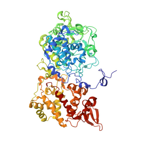
    Isoniazid (INH) is one of the most effective antibiotics against tuberculosis. INH is a prodrug that is activated by KatG. Although extensive studies have been performed in order to understand the mechanism of KatG, even the binding site of INH in KatG remains controversial. In this study, we determined the crystal structure of KatG from Synechococcus elongatus PCC7942 (SeKatG) in a complex with INH at 2.12-Å resolution. Three INH molecules were bound to the molecular surface. One INH molecule was bound at the entrance to the ε-edge side of heme (designated site 1), another was bound at the entrance to the γ-edge side of heme (site 2), and another was bound to the loop structures in front of the heme propionate side chain (site 3). All of the interactions between KatG and the bound INH seemed to be weak, being mediated mainly by van der Waals contacts. Structural comparisons revealed that the identity and configuration of the residues in site 1 were very similar among SeKatG, Burkholderia pseudomallei KatG, and Mycobacterium tuberculosis KatG. In contrast, sites 2 and 3 were structurally diverse among the three proteins. Thus, site 1 is probably the common KatG INH-binding site. A static enzymatic analysis and thermal shift assay suggested that the INH-activating reaction does not proceed in site 1, but rather that this site may function as an initial trapping site for the INH molecule. Database: The atomic coordinates and structure factors have been deposited in the Protein Data Bank under the accession number 3WXO.


  • Organizational Affiliation

    Graduate School of Science, Osaka Prefecture University, Sakai, Osaka, Japan.


Macromolecules
Find similar proteins by:  (by identity cutoff)  |  3D Structure
Entity ID: 1
MoleculeChains Sequence LengthOrganismDetailsImage
Catalase-peroxidase710Synechococcus elongatus PCC 7942 = FACHB-805Mutation(s): 0 
Gene Names: katGSynpcc7942_1656
EC: 1.11.1.21
UniProt
Find proteins for Q31MN3 (Synechococcus elongatus (strain ATCC 33912 / PCC 7942 / FACHB-805))
Explore Q31MN3 
Go to UniProtKB:  Q31MN3
Entity Groups  
Sequence Clusters30% Identity50% Identity70% Identity90% Identity95% Identity100% Identity
UniProt GroupQ31MN3
Sequence Annotations
Expand
  • Reference Sequence
Small Molecules
Ligands 3 Unique
IDChains Name / Formula / InChI Key2D Diagram3D Interactions
HEM
Query on HEM

Download Ideal Coordinates CCD File 
B [auth A]PROTOPORPHYRIN IX CONTAINING FE
C34 H32 Fe N4 O4
KABFMIBPWCXCRK-RGGAHWMASA-L
NIZ
Query on NIZ

Download Ideal Coordinates CCD File 
C [auth A],
D [auth A],
E [auth A]
pyridine-4-carbohydrazide
C6 H7 N3 O
QRXWMOHMRWLFEY-UHFFFAOYSA-N
NA
Query on NA

Download Ideal Coordinates CCD File 
F [auth A],
G [auth A],
H [auth A],
I [auth A]
SODIUM ION
Na
FKNQFGJONOIPTF-UHFFFAOYSA-N
Experimental Data & Validation

Experimental Data

  • Method: X-RAY DIFFRACTION
  • Resolution: 2.12 Å
  • R-Value Free: 0.197 
  • R-Value Work: 0.158 
  • R-Value Observed: 0.160 
  • Space Group: P 41 21 2
Unit Cell:
Length ( Å )Angle ( ˚ )
a = 108.654α = 90
b = 108.654β = 90
c = 202.891γ = 90
Software Package:
Software NamePurpose
SCALEPACKdata scaling
REFMACrefinement
PDB_EXTRACTdata extraction
HKL-2000data collection
HKL-2000data reduction
HKL-2000data scaling
MOLREPphasing

Structure Validation

View Full Validation Report


Ligand Structure Quality Assessment 


Entry History 

Deposition Data

Revision History  (Full details and data files)

  • Version 1.0: 2015-01-21
    Type: Initial release
  • Version 1.1: 2024-03-20
    Changes: Data collection, Database references, Derived calculations