3WWV | pdb_00003wwv

C-terminal domain of stomatin operon partner protein 1510-C from Pyrococcus horikoshii


Experimental Data Snapshot

  • Method: X-RAY DIFFRACTION
  • Resolution: 2.40 Å
  • R-Value Free: 
    0.258 (Depositor), 0.259 (DCC) 
  • R-Value Work: 
    0.221 (Depositor), 0.221 (DCC) 
  • R-Value Observed: 
    0.225 (Depositor) 

Starting Model: experimental
View more details

wwPDB Validation 3D Report Full Report

Validation slider image for 3WWV

This is version 1.3 of the entry. See complete history

Literature

Crystal structure of the stomatin operon partner protein from Pyrococcus horikoshii indicates the formation of a multimeric assembly

Yokoyama, H.Matsui, I.

(2014) FEBS Open Bio 4: 804-812

  • DOI: https://doi.org/10.1016/j.fob.2014.09.002
  • Primary Citation Related Structures: 
    3WWV

  • PubMed Abstract: 

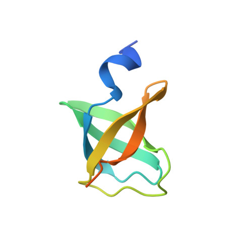
    Stomatin, prohibitin, flotillin, and HflK/C (SPFH) domain proteins are found in the lipid raft microdomains of various cellular membranes. Stomatin/STOPP (stomatin operon partner protein) gene pairs are present in both archaeal and bacterial species, and their protein products may be involved in the quality control of membrane proteins. In the present study, the crystal structure of the C-terminal soluble domain of STOPP PH1510 (1510-C) from the hyperthermophilic archaeon Pyrococcus horikoshii was determined at 2.4 Å resolution. The structure of 1510-C had a compact five-stranded β-barrel fold known as an oligosaccharide/oligonucleotide-binding fold (OB-fold). According to crystal packing, 1510-C could assemble into multimers based on a dimer as a basic unit. 1510-C also formed a large cylinder-like structure composed of 24 subunits or a large triangular prism-like structure composed of 12 subunits. These results indicate that 1510-C functions as a scaffold protein to form the multimeric assembly of STOPP and stomatin.


  • Organizational Affiliation
    • School of Pharmaceutical Sciences, University of Shizuoka, 52-1 Yada, Suruga-ku, Shizuoka 422-8526, Japan.

Macromolecule Content 

  • Total Structure Weight: 9.21 kDa 
  • Atom Count: 528 
  • Modeled Residue Count: 64 
  • Deposited Residue Count: 80 
  • Unique protein chains: 1

Macromolecules

Find similar proteins by:|  3D Structure
Entity ID: 1
MoleculeChains  Sequence LengthOrganismDetailsImage
Stomatin operon partner protein80Pyrococcus horikoshii OT3Mutation(s): 0 
Gene Names: PH1510
EC: 3.4.21
UniProt
Find proteins for O59179 (Pyrococcus horikoshii (strain ATCC 700860 / DSM 12428 / JCM 9974 / NBRC 100139 / OT-3))
Explore O59179 
Go to UniProtKB:  O59179
Entity Groups
Sequence Clusters30% Identity50% Identity70% Identity90% Identity95% Identity100% Identity
UniProt GroupO59179
Sequence Annotations
Expand
Reference Sequence

Small Molecules

Ligands 1 Unique
IDChains Name / Formula / InChI Key2D Diagram3D Interactions
NA

Query on NA


Download:Ideal Coordinates CCD File
B [auth A]SODIUM ION
Na
FKNQFGJONOIPTF-UHFFFAOYSA-N

Experimental Data & Validation

Experimental Data

  • Method: X-RAY DIFFRACTION
  • Resolution: 2.40 Å
  • R-Value Free:  0.258 (Depositor), 0.259 (DCC) 
  • R-Value Work:  0.221 (Depositor), 0.221 (DCC) 
  • R-Value Observed: 0.225 (Depositor) 
Space Group: P 62 2 2
Unit Cell:
Length ( Å )Angle ( ˚ )
a = 75.847α = 90
b = 75.847β = 90
c = 43.631γ = 120
Software Package:
Software NamePurpose
XDSdata scaling
PHENIXmodel building
REFMACrefinement
XDSdata reduction
SCALAdata scaling
PHENIXphasing

Structure Validation

View Full Validation Report


Entry History 

Deposition Data

Revision History  (Full details and data files)

  • Version 1.0: 2014-10-01
    Type: Initial release
  • Version 1.1: 2014-10-15
    Changes: Database references
  • Version 1.2: 2015-01-21
    Changes: Database references
  • Version 1.3: 2023-11-08
    Changes: Data collection, Database references, Derived calculations, Refinement description