3WV6

Crystal Structure of a protease-resistant mutant form of human galectin-9


Experimental Data Snapshot

  • Method: X-RAY DIFFRACTION
  • Resolution: 1.95 Å
  • R-Value Free: 0.222 
  • R-Value Work: 0.188 
  • R-Value Observed: 0.192 

wwPDB Validation   3D Report Full Report


This is version 2.0 of the entry. See complete history


Literature

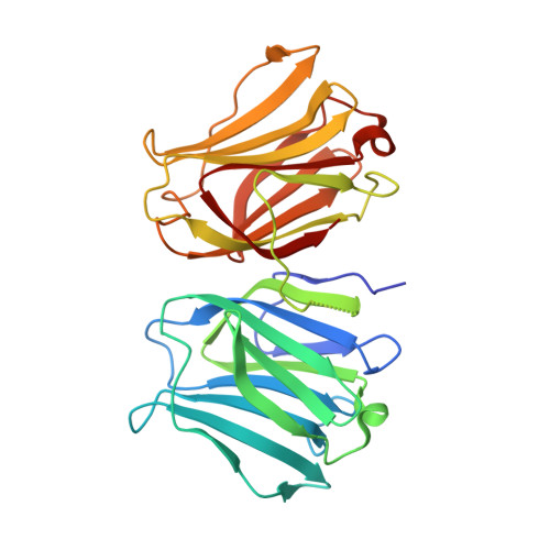
X-ray structure of a protease-resistant mutant form of human galectin-9 having two carbohydrate recognition domains with a metal-binding site

Yoshida, H.Nishi, N.Wada, K.Nakamura, T.Hirashima, M.Kuwabara, N.Kato, R.Kamitori, S.

(2017) Biochem Biophys Res Commun 490: 1287-1293

  • DOI: https://doi.org/10.1016/j.bbrc.2017.07.009
  • Primary Citation of Related Structures:  
    3WV6

  • PubMed Abstract: 
  • Galectin-9 (G9) is a tandem-repeat type β-galactoside-specific animal lectin having N-terminal and C-terminal carbohydrate recognition domains (N-CRD and C-CRD, respectively) joined by a linker peptide that is involved in the immune system. G9 is divalent in glycan binding, and structural information about the spatial arrangement of the two CRDs is very important for elucidating its biological functions ...

    Galectin-9 (G9) is a tandem-repeat type β-galactoside-specific animal lectin having N-terminal and C-terminal carbohydrate recognition domains (N-CRD and C-CRD, respectively) joined by a linker peptide that is involved in the immune system. G9 is divalent in glycan binding, and structural information about the spatial arrangement of the two CRDs is very important for elucidating its biological functions. As G9 is protease sensitive due to the long linker, the protease-resistant mutant form of G9 (G9Null) was developed by modification of the linker peptide, while retaining its biological functions. The X-ray structure of a mutant form of G9Null with the replacement of Arg221 by Ser (G9Null_R221S) having two CRDs was determined. The structure of G9Null_R221S was compact to associate the two CRDs in the back-to-back orientation with a large interface area, including hydrogen bonds and hydrophobic interactions. A metal ion was newly found in the galectin structure, possibly contributing to the stable structure of protein. The presented X-ray structure was thought to be one of the stable structures of G9, which likely occurs in solution. This was supported by structural comparisons with other tandem-repeated galectins and the analyses of protein thermostability by CD spectra measurements.


    Organizational Affiliation

    Life Science Research Center and Faculty of Medicine, Kagawa University, 1750-1, Ikenobe, Miki-cho, Kita-gun, Kagawa 761-0793, Japan. Electronic address: kamitori@med.kagawa-u.ac.jp.


Macromolecules
Find similar proteins by:  (by identity cutoff)  |  3D Structure
Entity ID: 1
MoleculeChainsSequence LengthOrganismDetailsImage
Galectin-9A, B296Homo sapiensMutation(s): 1 
Gene Names: LGALS9
UniProt & NIH Common Fund Data Resources
Find proteins for O00182 (Homo sapiens)
Explore O00182 
Go to UniProtKB:  O00182
PHAROS:  O00182
Entity Groups  
Sequence Clusters30% Identity50% Identity70% Identity90% Identity95% Identity100% Identity
UniProt GroupO00182
Protein Feature View
Expand
  • Reference Sequence
Oligosaccharides

Help

Entity ID: 2
MoleculeChainsChain Length2D DiagramGlycosylation3D Interactions
beta-D-galactopyranose-(1-4)-beta-D-glucopyranoseC, E 2N/A Oligosaccharides Interaction
Glycosylation Resources
GlyTouCan:  G84224TW
GlyCosmos:  G84224TW
GlyGen:  G84224TW
Entity ID: 3
MoleculeChainsChain Length2D DiagramGlycosylation3D Interactions
beta-D-galactopyranose-(1-4)-alpha-D-glucopyranoseD, F 2N/A Oligosaccharides Interaction
Glycosylation Resources
GlyTouCan:  G88362QR
GlyCosmos:  G88362QR
GlyGen:  G88362QR
Biologically Interesting Molecules (External Reference) 2 Unique
Entity ID: 2
IDChainsNameType/Class2D Diagram3D Interactions
PRD_900004
Query on PRD_900004
C, Ebeta-lactoseOligosaccharide / Nutrient Ligand Interaction
Entity ID: 3
IDChainsNameType/Class2D Diagram3D Interactions
PRD_900008
Query on PRD_900008
D, Falpha-lactoseOligosaccharide / Nutrient Ligand Interaction
Experimental Data & Validation

Experimental Data

  • Method: X-RAY DIFFRACTION
  • Resolution: 1.95 Å
  • R-Value Free: 0.222 
  • R-Value Work: 0.188 
  • R-Value Observed: 0.192 
  • Space Group: P 1 21 1
Unit Cell:
Length ( Å )Angle ( ˚ )
a = 58.34α = 90
b = 94.87β = 113.87
c = 64.81γ = 90
Software Package:
Software NamePurpose
HKL-2000data collection
MOLREPphasing
CNSrefinement
HKL-2000data reduction
HKL-2000data scaling

Structure Validation

View Full Validation Report


Entry History 

Deposition Data

Revision History  (Full details and data files)

  • Version 1.0: 2015-05-20
    Type: Initial release
  • Version 1.1: 2017-06-14
    Changes: Database references
  • Version 1.2: 2017-08-02
    Changes: Database references
  • Version 2.0: 2020-07-29
    Type: Remediation
    Reason: Carbohydrate remediation
    Changes: Atomic model, Data collection, Database references, Derived calculations, Structure summary