3WSE | pdb_00003wse

Reduced HcgD from Methanocaldococcus jannaschii


Experimental Data Snapshot

  • Method: X-RAY DIFFRACTION
  • Resolution: 2.50 Å
  • R-Value Free: 
    0.221 (Depositor), 0.227 (DCC) 
  • R-Value Work: 
    0.194 (Depositor), 0.201 (DCC) 
  • R-Value Observed: 
    0.196 (Depositor) 

Starting Model: experimental
View more details

wwPDB Validation 3D Report Full Report

Validation slider image for 3WSE

This is version 1.1 of the entry. See complete history

Literature

A possible iron delivery function of the dinuclear iron center of HcgD in [Fe]-hydrogenase cofactor biosynthesis

Fujishiro, T.Ermler, U.Shima, S.

(2014) FEBS Lett 588: 2789-2793

  • DOI: https://doi.org/10.1016/j.febslet.2014.05.059
  • Primary Citation Related Structures: 
    3WSD, 3WSE, 3WSF, 3WSG, 3WSH, 3WSI

  • PubMed Abstract: 

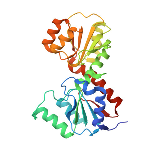
    HcgD, a homolog of the ubiquitous Nif3-like protein family, is found in a gene cluster involved in the biosynthesis of the iron-guanylylpyridinol (FeGP) cofactor of [Fe]-hydrogenase. The presented crystal structure and biochemical analyses indicated that HcgD has a dinuclear iron-center, which provides a pronounced binding site for anionic ligands. HcgD contains a stronger and a weaker bound iron; the latter being removable by chelating reagents preferentially in the oxidized state. Therefore, we propose HcgD as an iron chaperone in FeGP cofactor biosynthesis, which might also stimulate investigations on the functionally unknown but physiologically important eukaryotic Nif3-like protein family members.


  • Organizational Affiliation
    • Max Planck Institute for Terrestrial Microbiology, Karl-von-Frisch-Straße 10, 35043 Marburg, Germany.

Macromolecule Content 

  • Total Structure Weight: 170.77 kDa 
  • Atom Count: 11,973 
  • Modeled Residue Count: 1,473 
  • Deposited Residue Count: 1,512 
  • Unique protein chains: 1

Macromolecules

Find similar proteins by:|  3D Structure
Entity ID: 1
MoleculeChains  Sequence LengthOrganismDetailsImage
Putative GTP cyclohydrolase 1 type 2
A, B, C, D, E
A, B, C, D, E, F
252Methanocaldococcus jannaschii DSM 2661Mutation(s): 0 
Gene Names: MJ0927

Small Molecules

Ligands 2 Unique
IDChains Name / Formula / InChI Key2D Diagram3D Interactions
PO4

Query on PO4


Download:Ideal Coordinates CCD File
I [auth A]
L [auth B]
O [auth C]
R [auth D]
U [auth E]
I [auth A],
L [auth B],
O [auth C],
R [auth D],
U [auth E],
X [auth F]
PHOSPHATE ION
O4 P
NBIIXXVUZAFLBC-UHFFFAOYSA-K
FE2

Query on FE2


Download:Ideal Coordinates CCD File
G [auth A]
H [auth A]
J [auth B]
K [auth B]
M [auth C]
G [auth A],
H [auth A],
J [auth B],
K [auth B],
M [auth C],
N [auth C],
P [auth D],
Q [auth D],
S [auth E],
T [auth E],
V [auth F],
W [auth F]
FE (II) ION
Fe
CWYNVVGOOAEACU-UHFFFAOYSA-N

Experimental Data & Validation

Experimental Data

  • Method: X-RAY DIFFRACTION
  • Resolution: 2.50 Å
  • R-Value Free:  0.221 (Depositor), 0.227 (DCC) 
  • R-Value Work:  0.194 (Depositor), 0.201 (DCC) 
  • R-Value Observed: 0.196 (Depositor) 
Space Group: P 21 21 2
Unit Cell:
Length ( Å )Angle ( ˚ )
a = 95.01α = 90
b = 134.98β = 90
c = 161.62γ = 90
Software Package:
Software NamePurpose
XSCALEdata scaling
EPMRphasing
PHENIXrefinement
PDB_EXTRACTdata extraction
XDSdata scaling

Structure Validation

View Full Validation Report


Entry History 

Deposition Data

Revision History  (Full details and data files)

  • Version 1.0: 2014-08-27
    Type: Initial release
  • Version 1.1: 2023-11-08
    Changes: Data collection, Database references, Derived calculations, Refinement description