3WRP | pdb_00003wrp

FLEXIBILITY OF THE DNA-BINDING DOMAINS OF TRP REPRESSOR


Experimental Data Snapshot

  • Method: X-RAY DIFFRACTION
  • Resolution: 1.80 Å
  • R-Value Observed: 
    0.204 (Depositor) 

wwPDB Validation 3D Report Full Report

Validation slider image for 3WRP

This is version 1.3 of the entry. See complete history

Literature

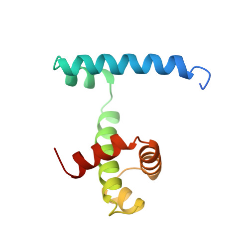
Flexibility of the DNA-binding domains of trp repressor.

Lawson, C.L.Zhang, R.G.Schevitz, R.W.Otwinowski, Z.Joachimiak, A.Sigler, P.B.

(1988) Proteins 3: 18-31

  • DOI: https://doi.org/10.1002/prot.340030103
  • Primary Citation Related Structures: 
    1WRP, 2OZ9, 3WRP

  • PubMed Abstract: 

    An orthorhombic crystal form of trp repressor (aporepressor plus L-tryptophan ligand) was solved by molecular replacement, refined to 1.65 A resolution, and compared to the structure of the repressor in trigonal crystals. Even though these two crystal forms of repressor were grown under identical conditions, the refined structures have distinctly different conformations of the DNA-binding domains. Unlike the repressor/aporepressor structural transition, the conformational shift is not caused by the binding or loss of the L-tryptophan ligand. We conclude that while L-tryptophan binding is essential for forming a specific complex with trp operator DNA, the corepressor ligand does not lock the repressor into a single conformation that is complementary to the operator. This flexibility may be required by the various binding modes proposed for trp repressor in its search for and adherence to its three different operator sites.


  • Organizational Affiliation
    • Department of Biochemistry and Molecular Biology, University of Chicago, Illinois 60637.

Macromolecule Content 

  • Total Structure Weight: 12.37 kDa 
  • Atom Count: 884 
  • Modeled Residue Count: 101 
  • Deposited Residue Count: 108 
  • Unique protein chains: 1

Macromolecules

Find similar proteins by:|  3D Structure
Entity ID: 1
MoleculeChains  Sequence LengthOrganismDetailsImage
TRP REPRESSOR108Escherichia coliMutation(s): 0 
UniProt
Find proteins for P0A881 (Escherichia coli (strain K12))
Explore P0A881 
Go to UniProtKB:  P0A881
Entity Groups
Sequence Clusters30% Identity50% Identity70% Identity90% Identity95% Identity100% Identity
UniProt GroupP0A881
Sequence Annotations
Expand
Reference Sequence

Experimental Data & Validation

Experimental Data

  • Method: X-RAY DIFFRACTION
  • Resolution: 1.80 Å
  • R-Value Observed: 0.204 (Depositor) 
Space Group: P 21 2 21
Unit Cell:
Length ( Å )Angle ( ˚ )
a = 57.4α = 90
b = 34.2β = 90
c = 44.5γ = 90
Software Package:
Software NamePurpose
PROLSQrefinement

Structure Validation

View Full Validation Report


Entry History 

Deposition Data

Revision History  (Full details and data files)

  • Version 1.0: 1988-04-16
    Type: Initial release
  • Version 1.1: 2008-03-03
    Changes: Version format compliance
  • Version 1.2: 2011-07-13
    Changes: Derived calculations, Version format compliance
  • Version 1.3: 2024-02-28
    Changes: Data collection, Database references, Other