C(sp3)-H bond hydroxylation catalyzed by myoglobin reconstituted with manganese porphycene.
Oohora, K., Kihira, Y., Mizohata, E., Inoue, T., Hayashi, T.(2013) J Am Chem Soc 135: 17282-17285
- PubMed: 24191678 Search on PubMed
- DOI: https://doi.org/10.1021/ja409404k
- Primary Citation Related Structures:
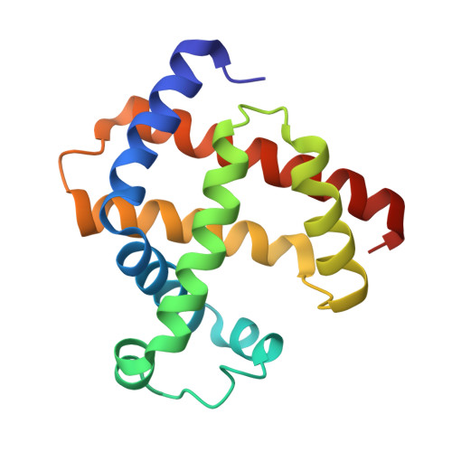
3WI8 - PubMed Abstract:
Myoglobin reconstituted with manganese porphycene was prepared in an effort to generate a new biocatalyst and was characterized by spectroscopic techniques. The X-ray crystal structure of the reconstituted protein reveals that the artificial cofactor is located in the intrinsic heme-binding site with weak ligation by His93. Interestingly, the reconstituted protein catalyzes the H2O2-dependent hydroxylation of ethylbenzene to yield 1-phenylethanol as a single product with a turnover number of 13 at 25 °C and pH 8.5. Native myoglobin and other modified myoglobins do not catalyze C-H hydroxylation of alkanes. Isotope effect experiments yield KIE values of 2.4 and 6.1 for ethylbenzene and toluene, respectively. Kinetic data, log kobs versus BDE(C(sp(3))-H) for ethylbenzene, toluene, and cyclohexane, indicate a linear relationship with a negative slope. These findings clearly indicate that the reaction occurs via a rate-determining step that involves hydrogen-atom abstraction by a Mn(O) species and a subsequent rebound hydroxylation process which is similar to the reaction mechanism of cytochrome P450.
- Department of Applied Chemistry, Graduate School of Engineering, Osaka University , Suita, 565-0871, Japan.
Organizational Affiliation: