3WFO | pdb_00003wfo

tRNA processing enzyme (apo form 1)


Experimental Data Snapshot

  • Method: X-RAY DIFFRACTION
  • Resolution: 3.40 Å
  • R-Value Free: 
    0.210 (Depositor), 0.208 (DCC) 
  • R-Value Work: 
    0.180 (Depositor), 0.182 (DCC) 
  • R-Value Observed: 
    0.181 (Depositor) 

wwPDB Validation 3D Report Full Report

Validation slider image for 3WFO

This is version 1.2 of the entry. See complete history

Literature

Translocation and rotation of tRNA during template-independent RNA polymerization by tRNA nucleotidyltransferase

Yamashita, S.Takeshita, D.Tomita, K.

(2014) Structure 22: 315-325

  • DOI: https://doi.org/10.1016/j.str.2013.12.002
  • Primary Citation Related Structures: 
    3WFO, 3WFP, 3WFQ, 3WFR, 3WFS

  • PubMed Abstract: 

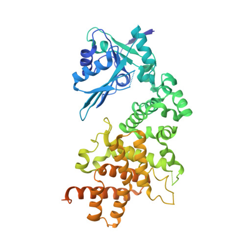
    The 3'-terminal CCA (CCA-3' at positions 74-76) of tRNA is synthesized by CCA-adding enzyme using CTP and ATP as substrates, without a nucleic acid template. In Aquifex aeolicus, CC-adding and A-adding enzymes collaboratively synthesize the CCA-3'. The mechanism of CCA-3' synthesis by these two enzymes remained obscure. We now present crystal structures representing CC addition onto tRNA by A. aeolicus CC-adding enzyme. After C₇₄ addition in an enclosed active pocket and pyrophosphate release, the tRNA translocates and rotates relative to the enzyme, and C₇₅ addition occurs in the same active pocket as C₇₄ addition. At both the C₇₄-adding and C₇₅-adding stages, CTP is selected by Watson-Crick-like hydrogen bonds between the cytosine of CTP and conserved Asp and Arg residues in the pocket. After C₇₄C₇₅ addition and pyrophosphate release, the tRNA translocates further and drops off the enzyme, and the CC-adding enzyme terminates RNA polymerization.


  • Organizational Affiliation
    • Biomedical Research Institute, National Institute of Advanced Industrial Science and Technology (AIST), 1-1-1, Higashi, Tsukuba, Ibaraki 305-8566, Japan.

Macromolecule Content 

  • Total Structure Weight: 182.65 kDa 
  • Atom Count: 10,470 
  • Modeled Residue Count: 1,276 
  • Deposited Residue Count: 1,575 
  • Unique protein chains: 1

Macromolecules

Find similar proteins by:|  3D Structure
Entity ID: 1
MoleculeChains  Sequence LengthOrganismDetailsImage
Poly A polymerase
A, B, C
525Aquifex aeolicus VF5Mutation(s): 0 
Gene Names: pcnB2
EC: 2.7.7.72 (PDB Primary Data), 2.7.7 (UniProt)
UniProt
Find proteins for O67911 (Aquifex aeolicus (strain VF5))
Explore O67911 
Go to UniProtKB:  O67911
Entity Groups
Sequence Clusters30% Identity50% Identity70% Identity90% Identity95% Identity100% Identity
UniProt GroupO67911
Sequence Annotations
Expand
Reference Sequence

Small Molecules

Ligands 1 Unique
IDChains Name / Formula / InChI Key2D Diagram3D Interactions
SO4

Query on SO4


Download:Ideal Coordinates CCD File
D [auth A]
E [auth A]
F [auth A]
G [auth A]
H [auth B]
D [auth A],
E [auth A],
F [auth A],
G [auth A],
H [auth B],
I [auth B],
J [auth B],
K [auth B],
L [auth C],
M [auth C],
N [auth C]
SULFATE ION
O4 S
QAOWNCQODCNURD-UHFFFAOYSA-L

Experimental Data & Validation

Experimental Data

  • Method: X-RAY DIFFRACTION
  • Resolution: 3.40 Å
  • R-Value Free:  0.210 (Depositor), 0.208 (DCC) 
  • R-Value Work:  0.180 (Depositor), 0.182 (DCC) 
  • R-Value Observed: 0.181 (Depositor) 
Space Group: C 2 2 2
Unit Cell:
Length ( Å )Angle ( ˚ )
a = 177.481α = 90
b = 327.769β = 90
c = 78.102γ = 90
Software Package:
Software NamePurpose
XDSdata scaling
PHASERphasing
PHENIXrefinement
XDSdata reduction

Structure Validation

View Full Validation Report


Entry History 

Deposition Data

Revision History  (Full details and data files)

  • Version 1.0: 2014-01-01
    Type: Initial release
  • Version 1.1: 2014-08-13
    Changes: Database references
  • Version 1.2: 2024-03-20
    Changes: Data collection, Database references, Derived calculations