3WDB | pdb_00003wdb

N-terminal domain of Mycobacterium tuberculosis ClpC1


Experimental Data Snapshot

  • Method: X-RAY DIFFRACTION
  • Resolution: 1.37 Å
  • R-Value Free: 
    0.182 (Depositor), 0.178 (DCC) 
  • R-Value Work: 
    0.143 (Depositor), 0.139 (DCC) 
  • R-Value Observed: 
    0.145 (Depositor) 

Starting Model: experimental
View more details

wwPDB Validation 3D Report Full Report

Validation slider image for 3WDB

This is version 1.2 of the entry. See complete history

Literature

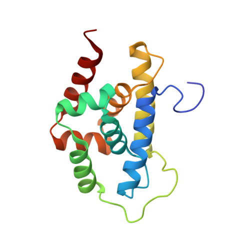
Structural basis of mycobacterial inhibition by cyclomarin A

Vasudevan, D.Rao, S.P.S.Noble, C.G.

(2013) J Biological Chem 288: 30883-30891

  • DOI: https://doi.org/10.1074/jbc.M113.493767
  • Primary Citation Related Structures: 
    3WDB, 3WDC, 3WDD, 3WDE

  • PubMed Abstract: 

    Cyclomarin A (CymA) was identified as a mycobactericidal compound targeting ClpC1. However, the target was identified based on pulldown experiments and in vitro binding data, without direct functional evidence in mycobacteria. Here we show that CymA specifically binds to the N-terminal domain of ClpC1. In addition we have determined the co-crystal structure of CymA bound to the N-terminal domain of ClpC1 to high resolution. Based on the structure of the complex several mutations were engineered into ClpC1, which showed reduced CymA binding in vitro. The ClpC1 mutants were overexpressed in mycobacteria and two showed resistance to CymA, providing the first direct evidence that ClpC1 is the target of CymA. Phe(80) is important in vitro and in cells for the ClpC1-CymA interaction and this explains why other bacteria are resistant to CymA. A model for how CymA binding to the N-terminal domain of ClpC1 leads to uncontrolled proteolysis by the associated ClpP protease machinery is discussed.


  • Organizational Affiliation
    • From the Novartis Institute for Tropical Diseases, 05-01 Chromos, Singapore 138670.

Macromolecule Content 

  • Total Structure Weight: 16.46 kDa 
  • Atom Count: 1,391 
  • Modeled Residue Count: 149 
  • Deposited Residue Count: 150 
  • Unique protein chains: 1

Macromolecules

Find similar proteins by:|  3D Structure
Entity ID: 1
MoleculeChains  Sequence LengthOrganismDetailsImage
Probable ATP-dependent Clp protease ATP-binding subunit150Mycobacterium tuberculosisMutation(s): 0 
Gene Names: clpC
UniProt
Find proteins for P9WPC9 (Mycobacterium tuberculosis (strain ATCC 25618 / H37Rv))
Explore P9WPC9 
Go to UniProtKB:  P9WPC9
Entity Groups
Sequence Clusters30% Identity50% Identity70% Identity90% Identity95% Identity100% Identity
UniProt GroupP9WPC9
Sequence Annotations
Expand
Reference Sequence

Small Molecules

Ligands 1 Unique
IDChains Name / Formula / InChI Key2D Diagram3D Interactions
FMT

Query on FMT


Download:Ideal Coordinates CCD File
B [auth A]FORMIC ACID
C H2 O2
BDAGIHXWWSANSR-UHFFFAOYSA-N

Experimental Data & Validation

Experimental Data

  • Method: X-RAY DIFFRACTION
  • Resolution: 1.37 Å
  • R-Value Free:  0.182 (Depositor), 0.178 (DCC) 
  • R-Value Work:  0.143 (Depositor), 0.139 (DCC) 
  • R-Value Observed: 0.145 (Depositor) 
Space Group: P 21 21 21
Unit Cell:
Length ( Å )Angle ( ˚ )
a = 42.37α = 90
b = 57.15β = 90
c = 65.02γ = 90
Software Package:
Software NamePurpose
MOLREPphasing
REFMACrefinement
MOSFLMdata reduction
SCALAdata scaling

Structure Validation

View Full Validation Report


Entry History 

Deposition Data

Revision History  (Full details and data files)

  • Version 1.0: 2013-09-18
    Type: Initial release
  • Version 1.1: 2013-12-11
    Changes: Database references
  • Version 1.2: 2023-11-08
    Changes: Data collection, Database references, Derived calculations, Refinement description