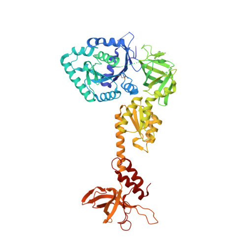
3WBK

crystal structure analysis of eukaryotic translation initiation factor 5B and 1A complex


Experimental Data Snapshot

  • Method: X-RAY DIFFRACTION
  • Resolution: 3.30 Å
  • R-Value Free: 0.317 
  • R-Value Work: 0.257 
  • R-Value Observed: 0.261 

Starting Model: experimental
View more details

wwPDB Validation   3D Report Full Report


This is version 1.3 of the entry. See complete history


Literature

X-ray structures of eIF5B and the eIF5B-eIF1A complex: the conformational flexibility of eIF5B is restricted on the ribosome by interaction with eIF1A

Zheng, A.Yu, J.Yamamoto, R.Ose, T.Tanaka, I.Yao, M.

(2014) Acta Crystallogr D Biol Crystallogr 70: 3090-3098

  • DOI: https://doi.org/10.1107/S1399004714021476
  • Primary Citation of Related Structures:  
    3WBI, 3WBJ, 3WBK

  • PubMed Abstract: 

    eIF5B and eIF1A are two translation-initiation factors that are universally conserved among all kingdoms. They show a unique interaction in eukaryotes which is important for ribosomal subunit joining. Here, the structures of two isolated forms of yeast eIF5B and of the eIF5B-eIF1A complex (eIF1A and eIF5B do not contain the respective N-terminal domains) are reported. The eIF5B-eIF1A structure shows that the C-terminal tail of eIF1A binds to eIF5B domain IV, while the core domain of eIF1A is invisible in the electron-density map. Although the individual domains in all structures of eIF5B or archaeal IF5B (aIF5B) are similar, their domain arrangements are significantly different, indicating high structural flexibility, which is advantageous for conformational change during ribosomal subunit joining. Based on these structures, models of eIF5B, eIF1A and tRNAi(Met) on the 80S ribosome were built. The models suggest that the interaction between the eIF1A C-terminal tail and eIF5B helps tRNAi(Met) to bind to eIF5B domain IV, thus preventing tRNAi(Met) dissociation, stabilizing the interface for subunit joining and providing a checkpoint for correct ribosome assembly.


  • Organizational Affiliation

    Graduate School of Life Sciences, Hokkaido University, Kita 10 Nishi 8 Kita-Ku, Sapporo, Hokkaido 060-0810, Japan.


Macromolecules
Find similar proteins by:  (by identity cutoff)  |  3D Structure
Entity ID: 1
MoleculeChains Sequence LengthOrganismDetailsImage
Eukaryotic translation initiation factor 5B
A, B
606Saccharomyces cerevisiae S288CMutation(s): 0 
Gene Names: FUN12YAL035W
UniProt
Find proteins for P39730 (Saccharomyces cerevisiae (strain ATCC 204508 / S288c))
Explore P39730 
Go to UniProtKB:  P39730
Entity Groups  
Sequence Clusters30% Identity50% Identity70% Identity90% Identity95% Identity100% Identity
UniProt GroupP39730
Sequence Annotations
Expand
  • Reference Sequence
Find similar proteins by:  (by identity cutoff)  |  3D Structure
Entity ID: 2
MoleculeChains Sequence LengthOrganismDetailsImage
Eukaryotic translation initiation factor 1A131Saccharomyces cerevisiae S288CMutation(s): 0 
Gene Names: TIF11YMR260CYM8156.02C
UniProt
Find proteins for P38912 (Saccharomyces cerevisiae (strain ATCC 204508 / S288c))
Explore P38912 
Go to UniProtKB:  P38912
Entity Groups  
Sequence Clusters30% Identity50% Identity70% Identity90% Identity95% Identity100% Identity
UniProt GroupP38912
Sequence Annotations
Expand
  • Reference Sequence
Experimental Data & Validation

Experimental Data

  • Method: X-RAY DIFFRACTION
  • Resolution: 3.30 Å
  • R-Value Free: 0.317 
  • R-Value Work: 0.257 
  • R-Value Observed: 0.261 
  • Space Group: P 21 21 21
Unit Cell:
Length ( Å )Angle ( ˚ )
a = 101.939α = 90
b = 120.942β = 90
c = 132.75γ = 90
Software Package:
Software NamePurpose
DENZOdata reduction
SCALEPACKdata scaling
REFMACrefinement
PDB_EXTRACTdata extraction
HKL-2000data reduction
HKL-2000data scaling
MOLREPphasing

Structure Validation

View Full Validation Report


Entry History 

Deposition Data

Revision History  (Full details and data files)

  • Version 1.0: 2014-11-19
    Type: Initial release
  • Version 1.1: 2015-02-04
    Changes: Database references
  • Version 1.2: 2017-11-22
    Changes: Refinement description
  • Version 1.3: 2023-11-08
    Changes: Data collection, Database references, Refinement description