3WBB

Crystal Structures of meso-diaminopimelate dehydrogenase from Symbiobacterium thermophilum


Experimental Data Snapshot

  • Method: X-RAY DIFFRACTION
  • Resolution: 1.93 Å
  • R-Value Free: 0.216 
  • R-Value Work: 0.179 

wwPDB Validation   3D Report Full Report


Ligand Structure Quality Assessment 


This is version 1.2 of the entry. See complete history


Literature

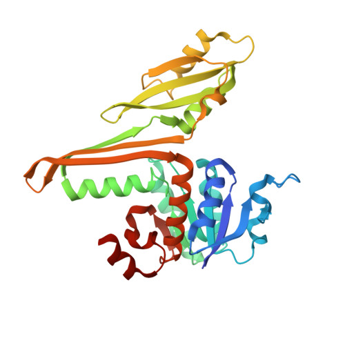
Structural and mutational studies on the unusual substrate specificity of meso-diaminopimelate dehydrogenase from Symbiobacterium thermophilum.

Liu, W.Li, Z.Huang, C.H.Guo, R.T.Zhao, L.Zhang, D.Chen, X.Wu, Q.Zhu, D.

(2014) Chembiochem 15: 217-222

  • DOI: https://doi.org/10.1002/cbic.201300691
  • Primary Citation of Related Structures:  
    3WB9, 3WBB, 3WBF

  • PubMed Abstract: 

    Wild-type meso-diaminopimelate dehydrogenase (DAPDH) is usually specific to the native substrate, meso-2,6-diaminopimelate. Recently, a DAPDH from Symbiobacterium thermophilum (StDAPDH) was found to exhibit expanded substrate specificity. As such, its crystal structures in apo form and in complex with NADP(+) and both NADPH and meso-DAP were investigated to reveal the structural basis of its unique catalytic properties. Structural analysis results show that StDAPDH should prefer an ordered kinetic catalytic mechanism. A second substrate entrance tunnel with Met152 at its bottleneck was found, through which pyruvate/D-alanine might bind and enter the catalytic cavity, providing some structural insights into its high activity toward pyruvate. The side chain of Met152 might interact with Asp92 and Asn253, thus affecting the domain motion and catalysis. These results offer useful information for understanding the unique catalytic properties of StDAPDH and guiding further engineering of this enzyme.


  • Organizational Affiliation

    Industrial Enzymes National Engineering Laboratory, Tianjin Institute of Industrial Biotechnology, Chinese Academy of Sciences, 32 Xi Qi Dao, Tianjin Airport Economic Area, Tianjin 300308 (China).


Macromolecules
Find similar proteins by:  (by identity cutoff)  |  3D Structure
Entity ID: 1
MoleculeChains Sequence LengthOrganismDetailsImage
Diaminopimelate dehydrogenase
A, B, C
305Symbiobacterium thermophilum IAM 14863Mutation(s): 0 
Gene Names: meso-dapdhSTH1425
EC: 1.4.1.16
UniProt
Find proteins for Q67PI3 (Symbiobacterium thermophilum (strain T / IAM 14863))
Explore Q67PI3 
Go to UniProtKB:  Q67PI3
Entity Groups  
Sequence Clusters30% Identity50% Identity70% Identity90% Identity95% Identity100% Identity
UniProt GroupQ67PI3
Sequence Annotations
Expand
  • Reference Sequence
Small Molecules
Ligands 2 Unique
IDChains Name / Formula / InChI Key2D Diagram3D Interactions
NAP
Query on NAP

Download Ideal Coordinates CCD File 
H [auth A],
L [auth B],
Q [auth C]
NADP NICOTINAMIDE-ADENINE-DINUCLEOTIDE PHOSPHATE
C21 H28 N7 O17 P3
XJLXINKUBYWONI-NNYOXOHSSA-N
GOL
Query on GOL

Download Ideal Coordinates CCD File 
D [auth A]
E [auth A]
F [auth A]
G [auth A]
I [auth B]
D [auth A],
E [auth A],
F [auth A],
G [auth A],
I [auth B],
J [auth B],
K [auth B],
M [auth C],
N [auth C],
O [auth C],
P [auth C]
GLYCEROL
C3 H8 O3
PEDCQBHIVMGVHV-UHFFFAOYSA-N
Experimental Data & Validation

Experimental Data

  • Method: X-RAY DIFFRACTION
  • Resolution: 1.93 Å
  • R-Value Free: 0.216 
  • R-Value Work: 0.179 
  • Space Group: C 1 2 1
Unit Cell:
Length ( Å )Angle ( ˚ )
a = 136.82α = 90
b = 99.95β = 112.25
c = 84.83γ = 90
Software Package:
Software NamePurpose
DENZOdata reduction
SCALEPACKdata scaling
MOLREPphasing
CNSrefinement
PDB_EXTRACTdata extraction
CrystalCleardata collection

Structure Validation

View Full Validation Report


Ligand Structure Quality Assessment 


Entry History 

Deposition Data

Revision History  (Full details and data files)

  • Version 1.0: 2014-03-26
    Type: Initial release
  • Version 1.1: 2017-11-22
    Changes: Refinement description
  • Version 1.2: 2023-11-08
    Changes: Data collection, Database references, Derived calculations, Refinement description