3WA0

Crystal structure of merlin complexed with DCAF1/VprBP


Experimental Data Snapshot

  • Method: X-RAY DIFFRACTION
  • Resolution: 2.31 Å
  • R-Value Free: 0.264 
  • R-Value Work: 0.235 
  • R-Value Observed: 0.237 

wwPDB Validation   3D Report Full Report


This is version 1.3 of the entry. See complete history


Literature

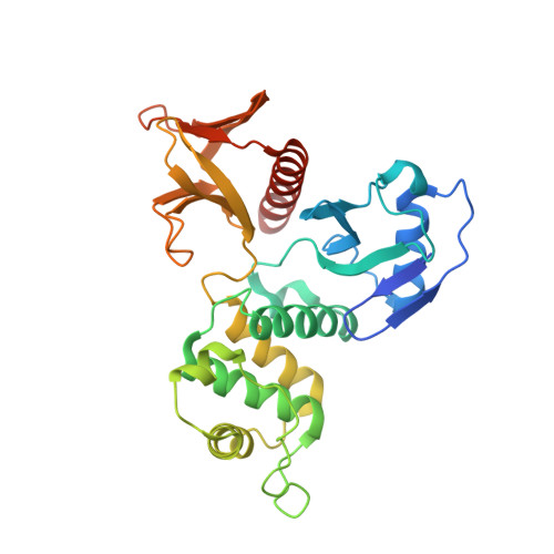
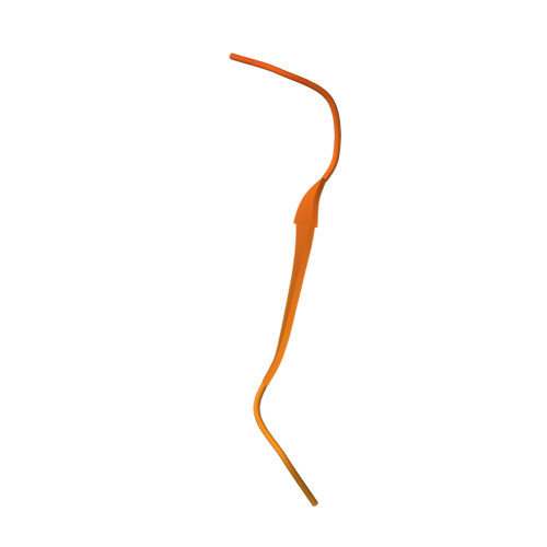
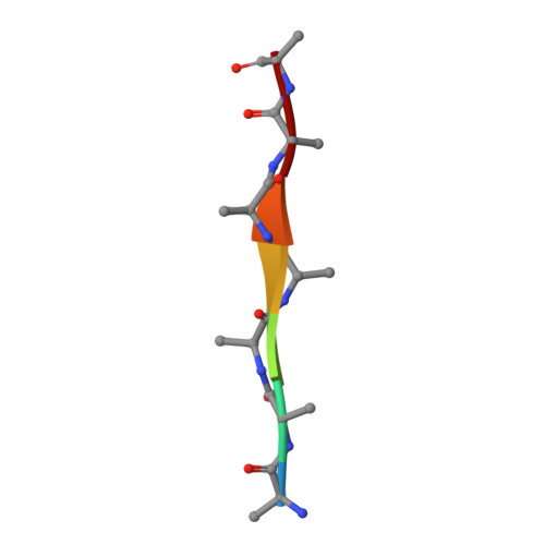
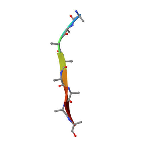
Structural basis of DDB1-and-Cullin 4-associated Factor 1 (DCAF1) recognition by merlin/NF2 and its implication in tumorigenesis by CD44-mediated inhibition of merlin suppression of DCAF1 function.

Mori, T.Gotoh, S.Shirakawa, M.Hakoshima, T.

(2014) Genes Cells 19: 603-619

  • DOI: https://doi.org/10.1111/gtc.12161
  • Primary Citation of Related Structures:  
    3WA0

  • PubMed Abstract: 

    Merlin, a tumor suppressor encoded by the neurofibromatosis type 2 gene, has been shown to suppress tumorigenesis by inhibiting the Cullin 4-RING E3 ubiquitin ligase CRL4(DCAF) (1) in the nucleus. This inhibition is mediated by direct binding of merlin to DDB1-and-Cullin 4-associated Factor 1 (DCAF1), yet the binding mode of merlin to DCAF1 is not well defined. Here, we report structural and biophysical studies of the merlin binding to DCAF1 and its interference with CD44 binding. The crystal structure of the merlin FERM domain bound to the DCAF1 C-terminal acidic tail reveals that the hydrophobic IILXLN motif located at the C-terminal end of DCAF1 binds subdomain C of the FERM domain by forming a β-strand. The binding site and mode resemble that of merlin binding to the CD44 cytoplasmic tail. Competition binding assay showed that CD44 and DCAF1 compete for binding to the merlin FERM domain in solution. The CD44 cytoplasmic tail is known to be cleaved for nuclear translocation by regulated intra-membrane proteolysis (RIP). Our structure implies that, in the nucleus, the CD44 cytoplasmic tail cleaved by RIP could release DCAF1 from merlin by competing for binding to the merlin FERM domain, which results in the inhibition of merlin-mediated suppression of tumorigenesis.


  • Organizational Affiliation

    Structural Biology Laboratory, Nara Institute of Science and Technology, 8916-5, Takayama, Ikoma, Nara, 630-0192, Japan.


Macromolecules
Find similar proteins by:  (by identity cutoff)  |  3D Structure
Entity ID: 1
MoleculeChains Sequence LengthOrganismDetailsImage
Merlin
A, B, C, D, E
A, B, C, D, E, F
301Mus musculusMutation(s): 0 
Gene Names: Nf2
UniProt
Find proteins for P46662 (Mus musculus)
Explore P46662 
Go to UniProtKB:  P46662
Entity Groups  
Sequence Clusters30% Identity50% Identity70% Identity90% Identity95% Identity100% Identity
UniProt GroupP46662
Sequence Annotations
Expand
  • Reference Sequence
Find similar proteins by:  (by identity cutoff)  |  3D Structure
Entity ID: 2
MoleculeChains Sequence LengthOrganismDetailsImage
Protein VPRBP
G, H
96Homo sapiensMutation(s): 0 
Gene Names: DCAF1
UniProt & NIH Common Fund Data Resources
Find proteins for Q9Y4B6 (Homo sapiens)
Explore Q9Y4B6 
Go to UniProtKB:  Q9Y4B6
PHAROS:  Q9Y4B6
GTEx:  ENSG00000145041 
Entity Groups  
Sequence Clusters30% Identity50% Identity70% Identity90% Identity95% Identity100% Identity
UniProt GroupQ9Y4B6
Sequence Annotations
Expand
  • Reference Sequence

Find similar proteins by:  Sequence   |   3D Structure  

Entity ID: 3
MoleculeChains Sequence LengthOrganismDetailsImage
Protein VPRBP7Homo sapiensMutation(s): 0 
Gene Names: DCAF1
Sequence Annotations
Expand
  • Reference Sequence

Find similar proteins by:  Sequence   |   3D Structure  

Entity ID: 4
MoleculeChains Sequence LengthOrganismDetailsImage
Protein VPRBP8Homo sapiensMutation(s): 0 
Gene Names: DCAF1
Sequence Annotations
Expand
  • Reference Sequence

Find similar proteins by:  Sequence   |   3D Structure  

Entity ID: 5
MoleculeChains Sequence LengthOrganismDetailsImage
Protein VPRBP6Homo sapiensMutation(s): 0 
Gene Names: DCAF1
Sequence Annotations
Expand
  • Reference Sequence
Experimental Data & Validation

Experimental Data

  • Method: X-RAY DIFFRACTION
  • Resolution: 2.31 Å
  • R-Value Free: 0.264 
  • R-Value Work: 0.235 
  • R-Value Observed: 0.237 
  • Space Group: P 21 21 21
Unit Cell:
Length ( Å )Angle ( ˚ )
a = 85.363α = 90
b = 135.76β = 90
c = 238.495γ = 90
Software Package:
Software NamePurpose
HKL-2000data collection
PHASERphasing
REFMACrefinement
HKL-2000data reduction
HKL-2000data scaling

Structure Validation

View Full Validation Report


Entry History 

Deposition Data

Revision History  (Full details and data files)

  • Version 1.0: 2014-05-28
    Type: Initial release
  • Version 1.1: 2014-06-25
    Changes: Database references, Structure summary
  • Version 1.2: 2022-08-24
    Changes: Database references
  • Version 1.3: 2023-11-08
    Changes: Data collection, Refinement description