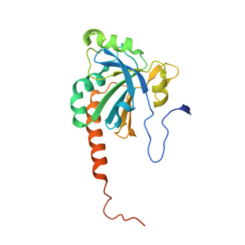
3VWV

crystal structure of N-terminally truncated peroxiredoxin 4 from M. musculus


Experimental Data Snapshot

  • Method: X-RAY DIFFRACTION
  • Resolution: 1.80 Å
  • R-Value Free: 0.193 
  • R-Value Work: 0.153 
  • R-Value Observed: 0.155 

wwPDB Validation   3D Report Full Report


This is version 1.0 of the entry. See complete history


Literature

Synergistic cooperation of PDI family members in peroxiredoxin 4-driven oxidative protein folding

Sato, Y.Kojima, R.Okumura, M.Hagiwara, M.Masui, S.Maegawa, K.Saiki, M.Horibe, T.Suzuki, M.Inaba, K.

(2013) Sci Rep 3: 2456-2456

  • DOI: https://doi.org/10.1038/srep02456
  • Primary Citation of Related Structures:  
    3VWU, 3VWV, 3VWW, 3W8J

  • PubMed Abstract: 

    The mammalian endoplasmic reticulum (ER) harbors disulfide bond-generating enzymes, including Ero1α and peroxiredoxin 4 (Prx4), and nearly 20 members of the protein disulfide isomerase family (PDIs), which together constitute a suitable environment for oxidative protein folding. Here, we clarified the Prx4 preferential recognition of two PDI family proteins, P5 and ERp46, and the mode of interaction between Prx4 and P5 thioredoxin domain. Detailed analyses of oxidative folding catalyzed by the reconstituted Prx4-PDIs pathways demonstrated that, while P5 and ERp46 are dedicated to rapid, but promiscuous, disulfide introduction, PDI is an efficient proofreader of non-native disulfides. Remarkably, the Prx4-dependent formation of native disulfide bonds was accelerated when PDI was combined with ERp46 or P5, suggesting that PDIs work synergistically to increase the rate and fidelity of oxidative protein folding. Thus, the mammalian ER seems to contain highly systematized oxidative networks for the efficient production of large quantities of secretory proteins.


  • Organizational Affiliation

    1] Division of Protein Chemistry, Medical Institute of Bioregulation, Kyushu University, 3-1-1 Maidashi, Higashi-ku, Fukuoka 812-8582, Japan [2].


Macromolecules
Find similar proteins by:  (by identity cutoff)  |  3D Structure
Entity ID: 1
MoleculeChains Sequence LengthOrganismDetailsImage
Peroxiredoxin-4
A, B
209Mus musculusMutation(s): 0 
Gene Names: Prdx4
EC: 1.11.1.15
UniProt
Find proteins for O08807 (Mus musculus)
Explore O08807 
Go to UniProtKB:  O08807
Entity Groups  
Sequence Clusters30% Identity50% Identity70% Identity90% Identity95% Identity100% Identity
UniProt GroupO08807
Sequence Annotations
Expand
  • Reference Sequence
Small Molecules
Ligands 2 Unique
IDChains Name / Formula / InChI Key2D Diagram3D Interactions
1PE
Query on 1PE

Download Ideal Coordinates CCD File 
C [auth A]PENTAETHYLENE GLYCOL
C10 H22 O6
JLFNLZLINWHATN-UHFFFAOYSA-N
ACY
Query on ACY

Download Ideal Coordinates CCD File 
D [auth A]ACETIC ACID
C2 H4 O2
QTBSBXVTEAMEQO-UHFFFAOYSA-N
Experimental Data & Validation

Experimental Data

  • Method: X-RAY DIFFRACTION
  • Resolution: 1.80 Å
  • R-Value Free: 0.193 
  • R-Value Work: 0.153 
  • R-Value Observed: 0.155 
  • Space Group: I 4
Unit Cell:
Length ( Å )Angle ( ˚ )
a = 104.321α = 90
b = 104.321β = 90
c = 84.228γ = 90
Software Package:
Software NamePurpose
BSSdata collection
PHASERphasing
PHENIXrefinement
HKL-2000data reduction
HKL-2000data scaling

Structure Validation

View Full Validation Report


Entry History 

Deposition Data

Revision History  (Full details and data files)

  • Version 1.0: 2013-09-04
    Type: Initial release