Structural insights into cellulolytic and chitinolytic enzymes revealing crucial residues of insect beta-N-acetyl-D-hexosaminidase
Liu, T., Zhou, Y., Chen, L., Chen, W., Liu, L., Shen, X., Zhang, W., Zhang, J., Yang, Q.(2012) PLoS One 7: e52225-e52225
- PubMed: 23300622 Search on PubMedSearch on PubMed Central
- DOI: https://doi.org/10.1371/journal.pone.0052225
- Primary Citation Related Structures:
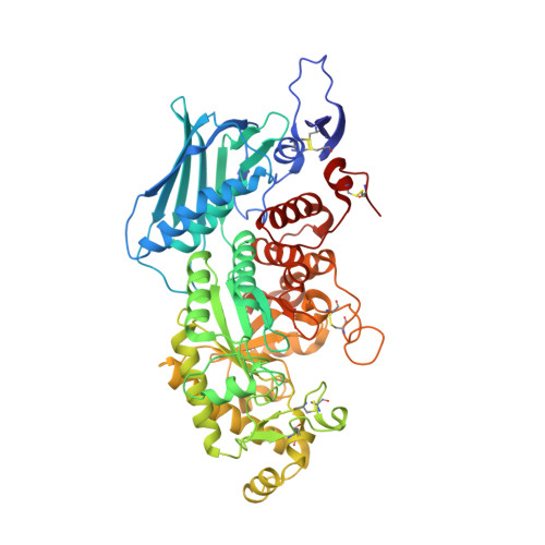
3VTR - PubMed Abstract:
The chemical similarity of cellulose and chitin supports the idea that their corresponding hydrolytic enzymes would bind β-1,4-linked glucose residues in a similar manner. A structural and mutational analysis was performed for the plant cellulolytic enzyme BGlu1 from Oryza sativa and the insect chitinolytic enzyme OfHex1 from Ostrinia furnacalis. Although BGlu1 shows little amino-acid sequence or topological similarity with OfHex1, three residues (Trp(490), Glu(328), Val(327) in OfHex1, and Trp(358), Tyr(131) and Ile(179) in BGlu1) were identified as being conserved in the +1 sugar binding site. OfHex1 Glu(328) together with Trp(490) was confirmed to be necessary for substrate binding. The mutant E328A exhibited a 8-fold increment in K(m) for (GlcNAc)(2) and a 42-fold increment in K(i) for TMG-chitotriomycin. A crystal structure of E328A in complex with TMG-chitotriomycin was resolved at 2.5 Å, revealing the obvious conformational changes of the catalytic residues (Glu(368) and Asp(367)) and the absence of the hydrogen bond between E328A and the C3-OH of the +1 sugar. V327G exhibited the same activity as the wild-type, but acquired the ability to efficiently hydrolyse β-1,2-linked GlcNAc in contrast to the wild-type. Thus, Glu(328) and Val(327) were identified as important for substrate-binding and as glycosidic-bond determinants. A structure-based sequence alignment confirmed the spatial conservation of these three residues in most plant cellulolytic, insect and bacterial chitinolytic enzymes.
- School of Life Science and Biotechnology, Dalian University of Technology, Dalian, China.
Organizational Affiliation: