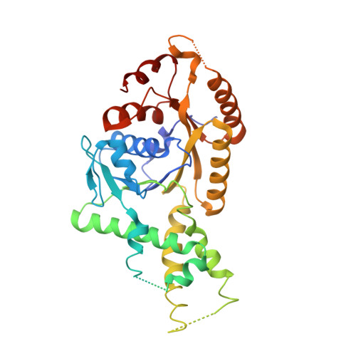
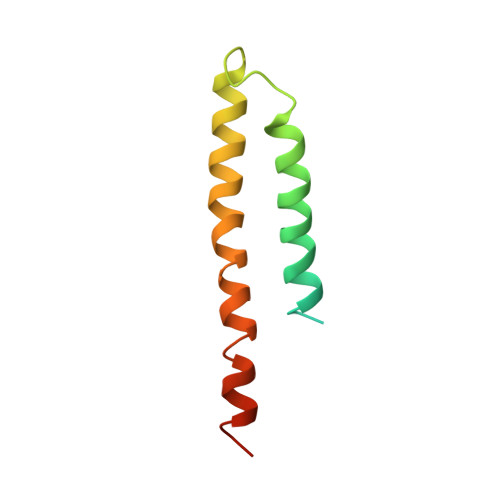
3VLC

Crystal structure of S. cerevisiae Get3 in the semi open conformation in complex with Get1 cytosolic domain at 4.5 angstrom resolution


Experimental Data Snapshot

  • Method: X-RAY DIFFRACTION
  • Resolution: 4.50 Å
  • R-Value Free: 0.366 
  • R-Value Work: 0.346 

Starting Model: experimental
View more details

wwPDB Validation   3D Report Full Report


Ligand Structure Quality Assessment 


This is version 1.2 of the entry. See complete history


Literature

Get1 stabilizes an open dimer conformation of get3 ATPase by binding two distinct interfaces

Kubota, K.Yamagata, A.Sato, Y.Goto-Ito, S.Fukai, S.

(2012) J Mol Biol 422: 366-375

  • DOI: https://doi.org/10.1016/j.jmb.2012.05.045
  • Primary Citation of Related Structures:  
    3B2E, 3VLC

  • PubMed Abstract: 

    Tail-anchored (TA) proteins are integral membrane proteins that possess a single transmembrane domain near their carboxy terminus. TA proteins play critical roles in many important cellular processes such as membrane trafficking, protein translocation, and apoptosis. The GET complex mediates posttranslational insertion of newly synthesized TA proteins to the endoplasmic reticulum membrane. The GET complex is composed of the homodimeric Get3 ATPase and its heterooligomeric receptor, Get1/2. During insertion, the Get3 dimer shuttles between open and closed conformational states, coupled with ATP hydrolysis and the binding/release of TA proteins. We report crystal structures of ADP-bound Get3 in complex with the cytoplasmic domain of Get1 (Get1CD) in open and semi-open conformations at 3.0- and 4.5-Å resolutions, respectively. Our structures and biochemical data suggest that Get1 uses two interfaces to stabilize the open dimer conformation of Get3. We propose that one interface is sufficient for binding of Get1 by Get3, while the second interface stabilizes the open dimer conformation of Get3.


  • Organizational Affiliation

    Structural Biology Laboratory, Life Science Division, Synchrotron Radiation Research Organization and Institute of Molecular and Cellular Biosciences, the University of Tokyo, Tokyo 113‐0032, Japan.


Macromolecules
Find similar proteins by:  (by identity cutoff)  |  3D Structure
Entity ID: 1
MoleculeChains Sequence LengthOrganismDetailsImage
ATPase GET3354Saccharomyces cerevisiaeMutation(s): 0 
EC: 3.6
UniProt
Find proteins for Q12154 (Saccharomyces cerevisiae (strain ATCC 204508 / S288c))
Explore Q12154 
Go to UniProtKB:  Q12154
Entity Groups  
Sequence Clusters30% Identity50% Identity70% Identity90% Identity95% Identity100% Identity
UniProt GroupQ12154
Sequence Annotations
Expand
  • Reference Sequence
Find similar proteins by:  (by identity cutoff)  |  3D Structure
Entity ID: 2
MoleculeChains Sequence LengthOrganismDetailsImage
Golgi to ER traffic protein 1B [auth E]94Saccharomyces cerevisiae S288CMutation(s): 0 
Gene Names: GET1MDM39YGL020C
UniProt
Find proteins for P53192 (Saccharomyces cerevisiae (strain ATCC 204508 / S288c))
Explore P53192 
Go to UniProtKB:  P53192
Entity Groups  
Sequence Clusters30% Identity50% Identity70% Identity90% Identity95% Identity100% Identity
UniProt GroupP53192
Sequence Annotations
Expand
  • Reference Sequence
Small Molecules
Ligands 1 Unique
IDChains Name / Formula / InChI Key2D Diagram3D Interactions
ADP
Query on ADP

Download Ideal Coordinates CCD File 
C [auth A]ADENOSINE-5'-DIPHOSPHATE
C10 H15 N5 O10 P2
XTWYTFMLZFPYCI-KQYNXXCUSA-N
Experimental Data & Validation

Experimental Data

  • Method: X-RAY DIFFRACTION
  • Resolution: 4.50 Å
  • R-Value Free: 0.366 
  • R-Value Work: 0.346 
  • Space Group: H 3 2
Unit Cell:
Length ( Å )Angle ( ˚ )
a = 132.634α = 90
b = 132.634β = 90
c = 185.153γ = 120
Software Package:
Software NamePurpose
HKL-2000data collection
MOLREPphasing
CNSrefinement
HKL-2000data reduction
HKL-2000data scaling

Structure Validation

View Full Validation Report


Ligand Structure Quality Assessment 


Entry History 

Deposition Data

Revision History  (Full details and data files)

  • Version 1.0: 2012-06-20
    Type: Initial release
  • Version 1.1: 2013-06-19
    Changes: Database references
  • Version 1.2: 2023-11-08
    Changes: Data collection, Database references, Derived calculations, Refinement description