3VHZ

Crystal structure of the trans isomer of the L93A mutant of bacteriorhodopsin


Experimental Data Snapshot

  • Method: X-RAY DIFFRACTION
  • Resolution: 2.30 Å
  • R-Value Free: 0.284 
  • R-Value Observed: 0.252 

wwPDB Validation   3D Report Full Report


Ligand Structure Quality Assessment 


This is version 2.1 of the entry. See complete history


Literature

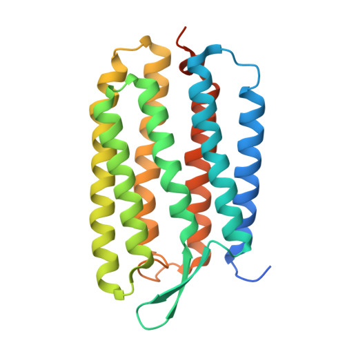
Crystal structure of the O intermediate of the Leu93Ala mutant of bacteriorhodopsin

Zhang, J.Yamazaki, Y.Hikake, M.Murakami, M.Ihara, K.Kouyama, T.

(2012) Proteins 80: 2384-2396

  • DOI: https://doi.org/10.1002/prot.24124
  • Primary Citation of Related Structures:  
    3VHZ, 3VI0

  • PubMed Abstract: 

    The lifetime of the O intermediate of bacteriorhodopsin (BR) is extended by a factor of ∼250 in the Leu93-to-Ala mutant (BR_L93A). To clarify the structural changes occurring in the last stage of the proton pumping cycle of BR, we crystallized BR_L93A into a hexagonal P622 crystal. Diffraction data from the unphotolyzed state showed that the deletion of three carbon atoms from Leu93 is compensated by the insertion of four water molecules in the cytoplasmic vicinity of retinal. This insertion of water is suggested to be responsible for the blue-shifted λ(max) (540 nm) of the mutant. A long-lived substate of O with a red-shifted λ(max) (~565 nm) was trapped when the crystal of BR_L93A was flash-cooled after illumination with green light. This substate (O(slow)) bears considerable similarity to the M intermediate of native BR; that is, it commonly shows deformation of helix C and the FG loop, downward orientation of the side chain of Arg82, and disruption of the Glu194/Glu204 pair. In O(slow), however, the main chain of Lys216 is less distorted and retinal takes on the 13-cis/15-syn configuration. Another significant difference is seen in the pH dependence of the structure of the proton release group, the pK(a) value of which is suggested to be much lower in O(slow) than in M.


  • Organizational Affiliation

    Department of Physics, Graduate School of Science, Nagoya University, Chikusa-Ku, Nagoya, Japan.


Macromolecules
Find similar proteins by:  (by identity cutoff)  |  3D Structure
Entity ID: 1
MoleculeChains Sequence LengthOrganismDetailsImage
Bacteriorhodopsin262Halobacterium salinarum NRC-1Mutation(s): 1 
Gene Names: bopVNG_1467G
Membrane Entity: Yes 
UniProt
Find proteins for P02945 (Halobacterium salinarum (strain ATCC 700922 / JCM 11081 / NRC-1))
Explore P02945 
Go to UniProtKB:  P02945
Entity Groups  
Sequence Clusters30% Identity50% Identity70% Identity90% Identity95% Identity100% Identity
UniProt GroupP02945
Sequence Annotations
Expand
  • Reference Sequence
Oligosaccharides

Help

Entity ID: 2
MoleculeChains Length2D Diagram Glycosylation3D Interactions
3-O-sulfo-beta-D-galactopyranose-(1-6)-alpha-D-mannopyranose-(1-2)-alpha-D-glucopyranose
B
3N/A
Glycosylation Resources
GlyTouCan:  G37414ZM
GlyCosmos:  G37414ZM
Small Molecules
Experimental Data & Validation

Experimental Data

  • Method: X-RAY DIFFRACTION
  • Resolution: 2.30 Å
  • R-Value Free: 0.284 
  • R-Value Observed: 0.252 
  • Space Group: P 6 2 2
Unit Cell:
Length ( Å )Angle ( ˚ )
a = 102.6α = 90
b = 102.6β = 90
c = 112.34γ = 120
Software Package:
Software NamePurpose
ADSCdata collection
CNSrefinement
MOSFLMdata reduction
SCALAdata scaling
CNSphasing

Structure Validation

View Full Validation Report


Ligand Structure Quality Assessment 


Entry History 

Deposition Data

Revision History  (Full details and data files)

  • Version 1.0: 2012-06-13
    Type: Initial release
  • Version 1.1: 2014-04-23
    Changes: Database references
  • Version 2.0: 2020-07-29
    Type: Remediation
    Reason: Carbohydrate remediation
    Changes: Atomic model, Data collection, Database references, Derived calculations, Structure summary
  • Version 2.1: 2023-11-08
    Changes: Data collection, Database references, Refinement description, Structure summary