3VFA | pdb_00003vfa

Crystal Structure of HIV-1 Protease Mutant V82A with novel P1'-Ligands GRL-02031


Experimental Data Snapshot

  • Method: X-RAY DIFFRACTION
  • Resolution: 1.43 Å
  • R-Value Free: 
    0.218 (Depositor), 0.205 (DCC) 
  • R-Value Work: 
    0.168 (Depositor), 0.165 (DCC) 
  • R-Value Observed: 
    0.170 (Depositor) 

Starting Model: experimental
View more details

wwPDB Validation 3D Report Full Report

Validation slider image for 3VFA

Ligand Structure Quality Assessment 


This is version 1.2 of the entry. See complete history

Literature

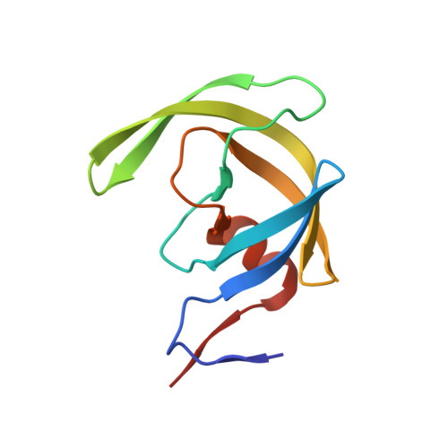
Potent antiviral HIV-1 protease inhibitor GRL-02031 adapts to the structures of drug resistant mutants with its P1'-pyrrolidinone ring.

Chang, Y.C.Yu, X.Zhang, Y.Tie, Y.Wang, Y.F.Yashchuk, S.Ghosh, A.K.Harrison, R.W.Weber, I.T.

(2012) J Med Chem 55: 3387-3397

  • DOI: https://doi.org/10.1021/jm300072d
  • Primary Citation Related Structures: 
    3VF5, 3VF7, 3VFA, 3VFB

  • PubMed Abstract: 

    GRL-02031 (1) is an HIV-1 protease (PR) inhibitor containing a novel P1' (R)-aminomethyl-2-pyrrolidinone group. Crystal structures at resolutions of 1.25-1.55 Å were analyzed for complexes of 1 with the PR containing major drug resistant mutations, PR(I47V), PR(L76V), PR(V82A), and PR(N88D). Mutations of I47V and V82A alter residues in the inhibitor-binding site, while L76V and N88D are distal mutations having no direct contact with the inhibitor. Substitution of a smaller amino acid in PR(I47V) and PR(L76V) and the altered charge of PR(N88D) are associated with significant local structural changes compared to the wild-type PR(WT), while substitution of alanine in PR(V82A) increases the size of the S1' subsite. The P1' pyrrolidinone group of 1 accommodates to these local changes by assuming two different conformations. Overall, the conformation and interactions of 1 with PR mutants resemble those of PR(WT) with similar inhibition constants in good agreement with the antiviral potency on multidrug resistant HIV-1.


  • Organizational Affiliation
    • Department of Biology, Molecular Basis of Disease Program, Georgia State University, P.O. Box 4010, Atlanta, Georgia 30302-4010, USA.

Macromolecule Content 

  • Total Structure Weight: 22.33 kDa 
  • Atom Count: 1,808 
  • Modeled Residue Count: 198 
  • Deposited Residue Count: 198 
  • Unique protein chains: 1

Macromolecules

Find similar proteins by:|  3D Structure
Entity ID: 1
MoleculeChains  Sequence LengthOrganismDetailsImage
protease
A, B
99Human immunodeficiency virus type 1 lw12.3 isolateMutation(s): 6 
Gene Names: gag-pol
EC: 3.4.23.16
UniProt
Find proteins for P0C6F2 (Human immunodeficiency virus type 1 group M subtype B (isolate LW123))
Explore P0C6F2 
Go to UniProtKB:  P0C6F2
Entity Groups
Sequence Clusters30% Identity50% Identity70% Identity90% Identity95% Identity100% Identity
UniProt GroupP0C6F2
Sequence Annotations
Expand
Reference Sequence

Small Molecules

Ligands 3 Unique
IDChains Name / Formula / InChI Key2D Diagram3D Interactions
031

Query on 031


Download:Ideal Coordinates CCD File
C [auth A](3aS,5R,6aR)-hexahydro-2H-cyclopenta[b]furan-5-yl [(1S,2R)-1-benzyl-2-hydroxy-3-([(4-methoxyphenyl)sulfonyl]{[(2R)-5-oxopyrrolidin-2-yl]methyl}amino)propyl]carbamate
C30 H39 N3 O8 S
RPIALZPTIFOQGC-CXLNPQPMSA-N
CL

Query on CL


Download:Ideal Coordinates CCD File
E [auth A]
F [auth A]
G [auth A]
H [auth A]
I [auth B]
E [auth A],
F [auth A],
G [auth A],
H [auth A],
I [auth B],
J [auth B],
K [auth B],
L [auth B]
CHLORIDE ION
Cl
VEXZGXHMUGYJMC-UHFFFAOYSA-M
NA

Query on NA


Download:Ideal Coordinates CCD File
D [auth A]SODIUM ION
Na
FKNQFGJONOIPTF-UHFFFAOYSA-N

Experimental Data & Validation

Experimental Data

  • Method: X-RAY DIFFRACTION
  • Resolution: 1.43 Å
  • R-Value Free:  0.218 (Depositor), 0.205 (DCC) 
  • R-Value Work:  0.168 (Depositor), 0.165 (DCC) 
  • R-Value Observed: 0.170 (Depositor) 
Space Group: P 21 21 2
Unit Cell:
Length ( Å )Angle ( ˚ )
a = 58.644α = 90
b = 86.459β = 90
c = 45.441γ = 90
Software Package:
Software NamePurpose
HKL-2000data collection
AMoREphasing
SHELXL-97refinement
HKL-2000data reduction
HKL-2000data scaling

Structure Validation

View Full Validation Report


Ligand Structure Quality Assessment 


Entry History 

Deposition Data

Revision History  (Full details and data files)

  • Version 1.0: 2012-11-21
    Type: Initial release
  • Version 1.1: 2015-03-25
    Changes: Database references
  • Version 1.2: 2023-09-13
    Changes: Data collection, Database references, Derived calculations, Refinement description