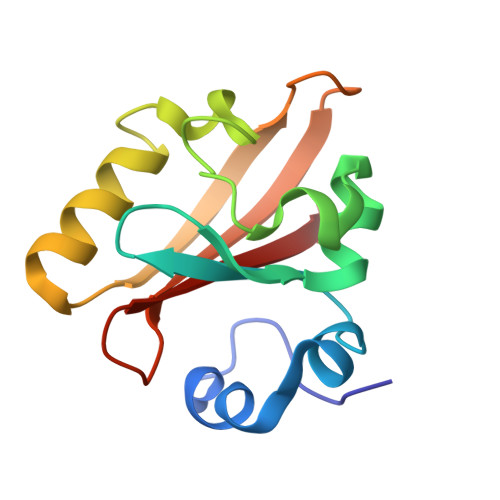
3VE3

Structure of IT Intermediate from time-resolved laue crystallography


Experimental Data Snapshot

  • Method: X-RAY DIFFRACTION
  • Resolution: 1.60 Å
  • R-Value Free: 0.291 
  • R-Value Observed: 0.239 

wwPDB Validation   3D Report Full Report


Ligand Structure Quality Assessment 


This is version 1.2 of the entry. See complete history


Literature

Volume-conserving trans-cis isomerization pathways in photoactive yellow protein visualized by picosecond X-ray crystallography

Jung, Y.O.Lee, J.H.Kim, J.Schmidt, M.Moffat, K.Srajer, V.Ihee, H.

(2013) Nat Chem 5: 212-220

  • DOI: https://doi.org/10.1038/nchem.1565
  • Primary Citation of Related Structures:  
    3VE3, 3VE4, 4HY8, 4I38, 4I39, 4I3A, 4I3I, 4I3J

  • PubMed Abstract: 

    Trans-to-cis isomerization, the key reaction in photoactive proteins, usually cannot occur through the standard one-bond-flip mechanism. Owing to spatial constraints imposed by a protein environment, isomerization probably proceeds through a volume-conserving mechanism in which highly choreographed atomic motions are expected, the details of which have not yet been observed directly. Here we employ time-resolved X-ray crystallography to visualize structurally the isomerization of the p-coumaric acid chromophore in photoactive yellow protein with a time resolution of 100 ps and a spatial resolution of 1.6 Å. The structure of the earliest intermediate (I(T)) resembles a highly strained transition state in which the torsion angle is located halfway between the trans- and cis-isomers. The reaction trajectory of I(T) bifurcates into two structurally distinct cis intermediates via hula-twist and bicycle-pedal pathways. The bifurcating reaction pathways can be controlled by weakening the hydrogen bond between the chromophore and an adjacent residue through E46Q mutation, which switches off the bicycle-pedal pathway.


  • Organizational Affiliation

    Center for Nanomaterials and Chemical Reactions, Institute for Basic Science, Daejeon 305-701, Republic of Korea.


Macromolecules
Find similar proteins by:  (by identity cutoff)  |  3D Structure
Entity ID: 1
MoleculeChains Sequence LengthOrganismDetailsImage
Photoactive yellow protein125Halorhodospira halophilaMutation(s): 0 
Gene Names: pyp
UniProt
Find proteins for P16113 (Halorhodospira halophila)
Explore P16113 
Go to UniProtKB:  P16113
Entity Groups  
Sequence Clusters30% Identity50% Identity70% Identity90% Identity95% Identity100% Identity
UniProt GroupP16113
Sequence Annotations
Expand
  • Reference Sequence
Small Molecules
Ligands 1 Unique
IDChains Name / Formula / InChI Key2D Diagram3D Interactions
HC4
Query on HC4

Download Ideal Coordinates CCD File 
B [auth A]4'-HYDROXYCINNAMIC ACID
C9 H8 O3
NGSWKAQJJWESNS-ZZXKWVIFSA-N
Experimental Data & Validation

Experimental Data

  • Method: X-RAY DIFFRACTION
  • Resolution: 1.60 Å
  • R-Value Free: 0.291 
  • R-Value Observed: 0.239 
  • Space Group: P 63
Unit Cell:
Length ( Å )Angle ( ˚ )
a = 66.833α = 90
b = 66.833β = 90
c = 40.947γ = 120
Software Package:
Software NamePurpose
LaueViewdata scaling
SHELXL-97refinement
LaueViewdata reduction

Structure Validation

View Full Validation Report


Ligand Structure Quality Assessment 


Entry History 

Deposition Data

Revision History  (Full details and data files)

  • Version 1.0: 2013-03-20
    Type: Initial release
  • Version 1.1: 2016-02-24
    Changes: Structure summary
  • Version 1.2: 2017-11-08
    Changes: Refinement description