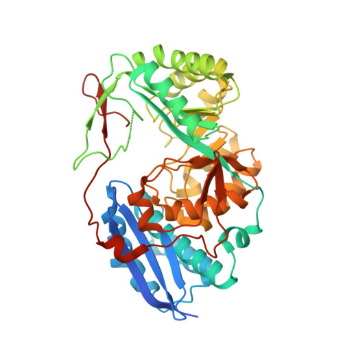
Crystal structure of an enolase from the soil bacterium Cellvibrio japonicus (TARGET EFI-502161) with bound MG and glycerol
Vetting, M.W., Toro, R., Bhosle, R., Wasserman, S.R., Morisco, L.L., Sojitra, S., Seidel, R., Hillerich, B., Washington, E., Scott Glenn, A., Chowdhury, S., Evans, B., Hammonds, J., Zencheck, W.D., Imker, H.J., Gerlt, J.A., Almo, S.C.To be published.