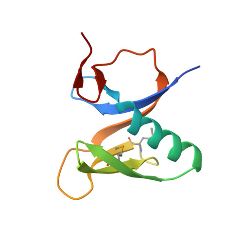
3V3L

Crystal structure of human RNF146 WWE domain in complex with iso-ADPRibose


Experimental Data Snapshot

  • Method: X-RAY DIFFRACTION
  • Resolution: 1.65 Å
  • R-Value Free: 0.181 
  • R-Value Work: 0.151 
  • R-Value Observed: 0.153 

wwPDB Validation   3D Report Full Report


Ligand Structure Quality Assessment 


This is version 1.0 of the entry. See complete history


Literature

Recognition of the iso-ADP-ribose moiety in poly(ADP-ribose) by WWE domains suggests a general mechanism for poly(ADP-ribosyl)ation-dependent ubiquitination.

Wang, Z.Michaud, G.A.Cheng, Z.Zhang, Y.Hinds, T.R.Fan, E.Cong, F.Xu, W.

(2012) Genes Dev 26: 235-240

  • DOI: https://doi.org/10.1101/gad.182618.111
  • Primary Citation of Related Structures:  
    3V3L

  • PubMed Abstract: 

    Protein poly(ADP-ribosyl)ation and ubiquitination are two key post-translational modifications regulating many biological processes. Through crystallographic and biochemical analysis, we show that the RNF146 WWE domain recognizes poly(ADP-ribose) (PAR) by interacting with iso-ADP-ribose (iso-ADPR), the smallest internal PAR structural unit containing the characteristic ribose-ribose glycosidic bond formed during poly(ADP-ribosyl)ation. The key iso-ADPR-binding residues we identified are highly conserved among WWE domains. Binding assays further demonstrate that PAR binding is a common function for the WWE domain family. Since many WWE domain-containing proteins are known E3 ubiquitin ligases, our results suggest that protein poly(ADP-ribosyl)ation may be a general mechanism to target proteins for ubiquitination.


  • Organizational Affiliation

    Department of Biological Structure, University of Washington, Seattle, Washington 98195, USA.


Macromolecules
Find similar proteins by:  (by identity cutoff)  |  3D Structure
Entity ID: 1
MoleculeChains Sequence LengthOrganismDetailsImage
E3 ubiquitin-protein ligase RNF146
A, B
85Homo sapiensMutation(s): 0 
Gene Names: RNF146
EC: 6.3.2
UniProt & NIH Common Fund Data Resources
Find proteins for Q9NTX7 (Homo sapiens)
Explore Q9NTX7 
Go to UniProtKB:  Q9NTX7
PHAROS:  Q9NTX7
GTEx:  ENSG00000118518 
Entity Groups  
Sequence Clusters30% Identity50% Identity70% Identity90% Identity95% Identity100% Identity
UniProt GroupQ9NTX7
Sequence Annotations
Expand
  • Reference Sequence
Small Molecules
Ligands 1 Unique
IDChains Name / Formula / InChI Key2D Diagram3D Interactions
V3L
Query on V3L

Download Ideal Coordinates CCD File 
C [auth A],
D [auth B]
2'-O-(5-O-phosphono-alpha-D-ribofuranosyl)adenosine 5'-(dihydrogen phosphate)
C15 H23 N5 O14 P2
BHIWBSNWEZIHHL-KEOHHSTQSA-N
Modified Residues  1 Unique
IDChains TypeFormula2D DiagramParent
MSE
Query on MSE
A, B
L-PEPTIDE LINKINGC5 H11 N O2 SeMET
Binding Affinity Annotations 
IDSourceBinding Affinity
V3L PDBBind:  3V3L Kd: 370 (nM) from 1 assay(s)
Binding MOAD:  3V3L Kd: 370 (nM) from 1 assay(s)
Experimental Data & Validation

Experimental Data

  • Method: X-RAY DIFFRACTION
  • Resolution: 1.65 Å
  • R-Value Free: 0.181 
  • R-Value Work: 0.151 
  • R-Value Observed: 0.153 
  • Space Group: P 31 1 2
Unit Cell:
Length ( Å )Angle ( ˚ )
a = 55.343α = 90
b = 55.343β = 90
c = 146.471γ = 120
Software Package:
Software NamePurpose
HKL-2000data collection
SOLVEphasing
REFMACrefinement
HKL-2000data reduction
HKL-2000data scaling

Structure Validation

View Full Validation Report


Ligand Structure Quality Assessment 


Entry History 

Deposition Data

Revision History  (Full details and data files)

  • Version 1.0: 2012-02-15
    Type: Initial release