3UL1

Mouse importin alpha: nucleoplasmin cNLS peptide complex


Experimental Data Snapshot

  • Method: X-RAY DIFFRACTION
  • Resolution: 1.90 Å
  • R-Value Free: 0.222 
  • R-Value Work: 0.191 
  • R-Value Observed: 0.193 

wwPDB Validation   3D Report Full Report


This is version 1.1 of the entry. See complete history


Literature

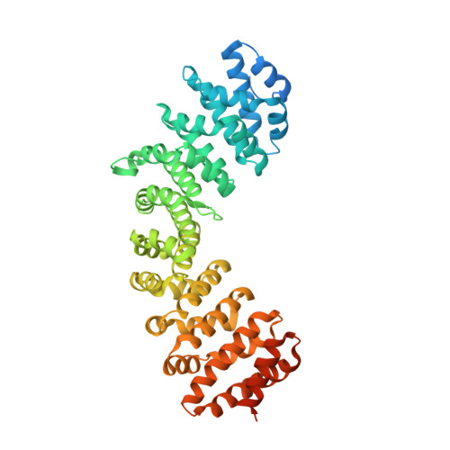
Structural Basis of High-Affinity Nuclear Localization Signal Interactions with Importin-alpha

Marfori, M.Lonhienne, T.G.Forwood, J.K.Kobe, B.

(2012) Traffic 13: 532-548

  • DOI: https://doi.org/10.1111/j.1600-0854.2012.01329.x
  • Primary Citation of Related Structures:  
    3UKW, 3UKX, 3UKY, 3UKZ, 3UL0, 3UL1

  • PubMed Abstract: 

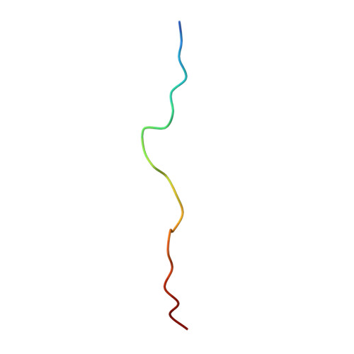
    Classical nuclear localization signals (cNLSs), comprising one (monopartite cNLSs) or two clusters of basic residues connected by a 10-12 residue linker (bipartite cNLSs), are recognized by the nuclear import factor importin-α. The cNLSs bind along a concave groove on importin-α; however, specificity determinants of cNLSs remain poorly understood. We present a structural and interaction analysis study of importin-α binding to both designed and naturally occurring high-affinity cNLS-like sequences; the peptide inhibitors Bimax1 and Bimax2, and cNLS peptides of cap-binding protein 80. Our data suggest that cNLSs and cNLS-like sequences can achieve high affinity through maximizing interactions at the importin-α minor site, and by taking advantage of multiple linker region interactions. Our study defines an extended set of binding cavities on the importin-α surface, and also expands on recent observations that longer linker sequences are allowed, and that long-range electrostatic complementarity can contribute to cNLS-binding affinity. Altogether, our study explains the molecular and structural basis of the results of a number of recent studies, including systematic mutagenesis and peptide library approaches, and provides an improved level of understanding on the specificity determinants of a cNLS. Our results have implications for identifying cNLSs in novel proteins.


  • Organizational Affiliation

    School of Chemistry and Molecular Biosciences, University of Queensland, Brisbane, QLD, 4072, Australia.


Macromolecules
Find similar proteins by:  (by identity cutoff)  |  3D Structure
Entity ID: 1
MoleculeChains Sequence LengthOrganismDetailsImage
Importin subunit alpha-2A [auth B]510Mus musculusMutation(s): 0 
Gene Names: Kpna2
UniProt & NIH Common Fund Data Resources
Find proteins for P52293 (Mus musculus)
Explore P52293 
Go to UniProtKB:  P52293
IMPC:  MGI:103561
Entity Groups  
Sequence Clusters30% Identity50% Identity70% Identity90% Identity95% Identity100% Identity
UniProt GroupP52293
Sequence Annotations
Expand
  • Reference Sequence

Find similar proteins by:  Sequence   |   3D Structure  

Entity ID: 2
MoleculeChains Sequence LengthOrganismDetailsImage
NucleoplasminB [auth A]22Xenopus laevisMutation(s): 0 
UniProt
Find proteins for P05221 (Xenopus laevis)
Explore P05221 
Go to UniProtKB:  P05221
Entity Groups  
Sequence Clusters30% Identity50% Identity70% Identity90% Identity95% Identity100% Identity
UniProt GroupP05221
Sequence Annotations
Expand
  • Reference Sequence
Experimental Data & Validation

Experimental Data

  • Method: X-RAY DIFFRACTION
  • Resolution: 1.90 Å
  • R-Value Free: 0.222 
  • R-Value Work: 0.191 
  • R-Value Observed: 0.193 
  • Space Group: P 21 21 21
Unit Cell:
Length ( Å )Angle ( ˚ )
a = 79.156α = 90
b = 90.094β = 90
c = 99.332γ = 90
Software Package:
Software NamePurpose
CrystalCleardata collection
PHENIXrefinement
d*TREKdata reduction
d*TREKdata scaling

Structure Validation

View Full Validation Report


Entry History 

Deposition Data

Revision History  (Full details and data files)

  • Version 1.0: 2012-10-03
    Type: Initial release
  • Version 1.1: 2023-11-01
    Changes: Data collection, Database references, Refinement description