3UIG

crystal structure of human Survivin in complex with T3 phosphorylated H3(1-15) peptide


Experimental Data Snapshot

  • Method: X-RAY DIFFRACTION
  • Resolution: 2.40 Å
  • R-Value Free: 0.250 
  • R-Value Work: 0.213 
  • R-Value Observed: 0.215 

wwPDB Validation   3D Report Full Report


This is version 1.3 of the entry. See complete history


Literature

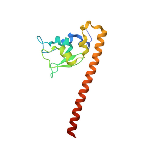
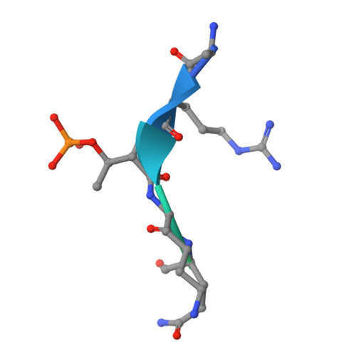
Structural Basis for Recognition of H3T3ph and Smac/DIABLO N-terminal Peptides by Human Survivin.

Du, J.Kelly, A.E.Funabiki, H.Patel, D.J.

(2012) Structure 20: 185-195

  • DOI: https://doi.org/10.1016/j.str.2011.12.001
  • Primary Citation of Related Structures:  
    3UIG, 3UIH, 3UII, 3UIJ, 3UIK

  • PubMed Abstract: 

    Survivin is an inhibitor of apoptosis family protein implicated in apoptosis and mitosis. In apoptosis, it has been shown to recognize the Smac/DIABLO protein. It is also a component of the chromosomal passenger complex, a key player during mitosis. Recently, Survivin was identified in vitro and in vivo as the direct binding partner for phosphorylated Thr3 on histone H3 (H3T3ph). We have undertaken structural and binding studies to investigate the molecular basis underlying recognition of H3T3ph and Smac/DIABLO N-terminal peptides by Survivin. Our crystallographic studies establish recognition of N-terminal Ala in both complexes and identify intermolecular hydrogen-bonding interactions in the Survivin phosphate-binding pocket that contribute to H3T3ph mark recognition. In addition, our calorimetric data establish that Survivin binds tighter to the H3T3ph-containing peptide relative to the N-terminal Smac/DIABLO peptide, and this preference can be reversed through structure-guided mutations that increase the hydrophobicity of the phosphate-binding pocket.


  • Organizational Affiliation

    Structural Biology Program, Memorial Sloan-Kettering Cancer Center, New York, NY 10065, USA.


Macromolecules
Find similar proteins by:  (by identity cutoff)  |  3D Structure
Entity ID: 1
MoleculeChains Sequence LengthOrganismDetailsImage
Baculoviral IAP repeat-containing protein 5
A, B
143Homo sapiensMutation(s): 1 
Gene Names: BIRC5API4IAP4
UniProt & NIH Common Fund Data Resources
Find proteins for O15392 (Homo sapiens)
Explore O15392 
Go to UniProtKB:  O15392
PHAROS:  O15392
GTEx:  ENSG00000089685 
Entity Groups  
Sequence Clusters30% Identity50% Identity70% Identity90% Identity95% Identity100% Identity
UniProt GroupO15392
Sequence Annotations
Expand
  • Reference Sequence

Find similar proteins by:  Sequence   |   3D Structure  

Entity ID: 2
MoleculeChains Sequence LengthOrganismDetailsImage
T3 phosphorylated H3(1-15) peptideC [auth P],
D [auth Q]
15Homo sapiensMutation(s): 0 
Gene Names: H3C1H3C2H3C3H3C4H3C6H3C7H3C8H3C10H3C11H3C12
UniProt & NIH Common Fund Data Resources
Find proteins for P68431 (Homo sapiens)
Explore P68431 
Go to UniProtKB:  P68431
PHAROS:  P68431
Entity Groups  
Sequence Clusters30% Identity50% Identity70% Identity90% Identity95% Identity100% Identity
UniProt GroupP68431
Sequence Annotations
Expand
  • Reference Sequence
Small Molecules
Modified Residues  1 Unique
IDChains TypeFormula2D DiagramParent
TPO
Query on TPO
C [auth P],
D [auth Q]
L-PEPTIDE LINKINGC4 H10 N O6 PTHR
Experimental Data & Validation

Experimental Data

  • Method: X-RAY DIFFRACTION
  • Resolution: 2.40 Å
  • R-Value Free: 0.250 
  • R-Value Work: 0.213 
  • R-Value Observed: 0.215 
  • Space Group: C 1 2 1
Unit Cell:
Length ( Å )Angle ( ˚ )
a = 115.778α = 90
b = 71.042β = 128.81
c = 82.287γ = 90
Software Package:
Software NamePurpose
ADSCdata collection
PHENIXmodel building
PHENIXrefinement
HKL-2000data reduction
HKL-2000data scaling
PHENIXphasing

Structure Validation

View Full Validation Report


Entry History 

Deposition Data

Revision History  (Full details and data files)

  • Version 1.0: 2012-02-01
    Type: Initial release
  • Version 1.1: 2012-12-12
    Changes: Other
  • Version 1.2: 2013-04-24
    Changes: Other
  • Version 1.3: 2023-09-13
    Changes: Data collection, Database references, Derived calculations, Refinement description