3UDZ

Inositol 1,3,4,5,6-pentakisphosphate 2-kinase from A. thaliana in complex with ADP and IP6.


Experimental Data Snapshot

  • Method: X-RAY DIFFRACTION
  • Resolution: 2.50 Å
  • R-Value Free: 0.260 
  • R-Value Work: 0.189 
  • R-Value Observed: 0.193 

wwPDB Validation   3D Report Full Report


Ligand Structure Quality Assessment 


This is version 1.3 of the entry. See complete history


Literature

Inositol phosphate-induced stabilization of inositol 1,3,4,5,6-pentakisphosphate 2-kinase and its role in substrate specificity.

Gosein, V.Leung, T.F.Krajden, O.Miller, G.J.

(2012) Protein Sci 21: 737-742

  • DOI: https://doi.org/10.1002/pro.2049
  • Primary Citation of Related Structures:  
    3UDS, 3UDT, 3UDZ

  • PubMed Abstract: 

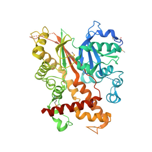
    Inositol phosphate kinases (IPKs) sequentially phosphorylate inositol phosphates (IPs) on their inositol rings to yield an array of signaling molecules. IPKs must possess the ability to recognize their physiological substrates from among a pool of over 30 cellular IPs that differ in numbers and positions of phosphates. Crystal structures from IPK subfamilies have revealed structural determinants for IP discrimination, which vary considerably between IPKs. However, recent structures of inositol 1,3,4,5,6-pentakisphosphate 2-kinase (IPK1) did not reveal how IPK1 selectively recognizes its physiological substrate, IP5, while excluding others. Here, we report that limited proteolysis has revealed the presence of multiple conformational states in the IPK1 catalytic cycle, with notable protection from protease only in the presence of IP. Further, a 3.1-Å crystal structure of IPK1 bound to ADP in the absence of IP revealed decreased order in residues 110-140 within the N-lobe of the kinase compared with structures in which IP is bound. Using this solution and crystallographic data, we propose a model for recognition of IP substrate by IPK1 wherein phosphate groups at the 4-, 5-, and 6-positions are recognized initially by the C-lobe with subsequent interaction of the 1-position phosphate by Arg130 that stabilizes this residue and the N-lobe. This model explains how IPK1 can be highly specific for a single IP substrate by linking its interactions with substrate phosphate groups to the stabilization of the N- and C-lobes and kinase activation.


  • Organizational Affiliation

    Department of Pharmacology and Therapeutics, McGill University, Montreal, Canada, H3G 1Y6.


Macromolecules
Find similar proteins by:  (by identity cutoff)  |  3D Structure
Entity ID: 1
MoleculeChains Sequence LengthOrganismDetailsImage
Inositol pentakisphosphate 2-kinase
A, B
493Arabidopsis thalianaMutation(s): 0 
Gene Names: At5g42810IPK1MJB21.19
EC: 2.7.1.158
UniProt
Find proteins for Q93YN9 (Arabidopsis thaliana)
Explore Q93YN9 
Go to UniProtKB:  Q93YN9
Entity Groups  
Sequence Clusters30% Identity50% Identity70% Identity90% Identity95% Identity100% Identity
UniProt GroupQ93YN9
Sequence Annotations
Expand
  • Reference Sequence
Small Molecules
Ligands 4 Unique
IDChains Name / Formula / InChI Key2D Diagram3D Interactions
IHP
Query on IHP

Download Ideal Coordinates CCD File 
C [auth A],
G [auth B]
INOSITOL HEXAKISPHOSPHATE
C6 H18 O24 P6
IMQLKJBTEOYOSI-GPIVLXJGSA-N
ADP
Query on ADP

Download Ideal Coordinates CCD File 
D [auth A],
H [auth B]
ADENOSINE-5'-DIPHOSPHATE
C10 H15 N5 O10 P2
XTWYTFMLZFPYCI-KQYNXXCUSA-N
ZN
Query on ZN

Download Ideal Coordinates CCD File 
K [auth B]ZINC ION
Zn
PTFCDOFLOPIGGS-UHFFFAOYSA-N
MG
Query on MG

Download Ideal Coordinates CCD File 
E [auth A],
F [auth A],
I [auth B],
J [auth B]
MAGNESIUM ION
Mg
JLVVSXFLKOJNIY-UHFFFAOYSA-N
Modified Residues  1 Unique
IDChains TypeFormula2D DiagramParent
MSE
Query on MSE
A, B
L-PEPTIDE LINKINGC5 H11 N O2 SeMET
Experimental Data & Validation

Experimental Data

  • Method: X-RAY DIFFRACTION
  • Resolution: 2.50 Å
  • R-Value Free: 0.260 
  • R-Value Work: 0.189 
  • R-Value Observed: 0.193 
  • Space Group: P 1
Unit Cell:
Length ( Å )Angle ( ˚ )
a = 59.057α = 89.84
b = 59.394β = 82.58
c = 82.755γ = 63.36
Software Package:
Software NamePurpose
DENZOdata reduction
SCALEPACKdata scaling
PHENIXrefinement
PDB_EXTRACTdata extraction
StructureStudiodata collection
HKL-2000data reduction
SOLVEphasing

Structure Validation

View Full Validation Report


Ligand Structure Quality Assessment 


Entry History 

Deposition Data

Revision History  (Full details and data files)

  • Version 1.0: 2012-03-14
    Type: Initial release
  • Version 1.1: 2012-05-09
    Changes: Database references
  • Version 1.2: 2017-11-08
    Changes: Refinement description
  • Version 1.3: 2020-10-14
    Changes: Database references, Derived calculations, Structure summary