Discovery and optimization of a novel spiropyrrolidine inhibitor of {beta}-secretase (BACE1) through fragment-based drug design.
Efremov, I.V., Vajdos, F.F., Borzilleri, K.A., Capetta, S., Chen, H., Dorff, P.H., Dutra, J.K., Goldstein, S.W., Mansour, M., McColl, A., Noell, S., Oborski, C.E., O'Connell, T.N., O'Sullivan, T.J., Pandit, J., Wang, H., Wei, B., Withka, J.M.(2012) J Med Chem 55: 9069-9088
- PubMed: 22468999 Search on PubMed
- DOI: https://doi.org/10.1021/jm201715d
- Primary Citation Related Structures:
3UDH, 3UDJ, 3UDK, 3UDM, 3UDN, 3UDP, 3UDQ, 3UDR, 3UDY - PubMed Abstract:
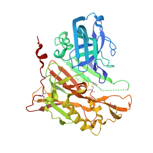
The aspartyl protease β-secretase, or BACE, has been demonstrated to be a key factor in the proteolytic formation of Aβ-peptide, a major component of plaques in the brains of Alzheimer's disease (AD) patients, and inhibition of this enzyme has emerged as a major strategy for pharmacologic intervention in AD. An X-ray-based fragment screen of Pfizer's proprietary fragment collection has resulted in the identification of a novel BACE binder featuring spiropyrrolidine framework. Although exhibiting only weak inhibitory activity against the BACE enzyme, the small compound was verified by biophysical and NMR-based methods as a bona fide BACE inhibitor. Subsequent optimization of the lead compound, relying heavily on structure-based drug design and computational prediction of physiochemical properties, resulted in a nearly 1000-fold improvement in potency while maintaining ligand efficiency and properties predictive of good permeability and low P-gp liability.
- Pfizer Worldwide Research, Groton Laboratories, Eastern Point Road, Groton, Connecticut 06340, United States. ivan.efremov@pfizer.com
Organizational Affiliation: