3U9S

Crystal structure of P. aeruginosa 3-methylcrotonyl-CoA carboxylase (MCC) 750 kD holoenzyme, CoA complex


Experimental Data Snapshot

  • Method: X-RAY DIFFRACTION
  • Resolution: 3.50 Å
  • R-Value Free: 0.292 
  • R-Value Work: 0.234 
  • R-Value Observed: 0.234 

wwPDB Validation   3D Report Full Report


Ligand Structure Quality Assessment 


This is version 1.1 of the entry. See complete history


Literature

An unanticipated architecture of the 750-kDa {alpha}6{beta}6 holoenzyme of 3-methylcrotonyl-CoA carboxylase

Huang, C.S.Ge, P.Zhou, Z.H.Tong, L.

(2012) Nature 481: 219-223

  • DOI: https://doi.org/10.1038/nature10691
  • Primary Citation of Related Structures:  
    3U9R, 3U9S, 3U9T

  • PubMed Abstract: 

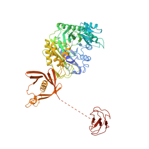
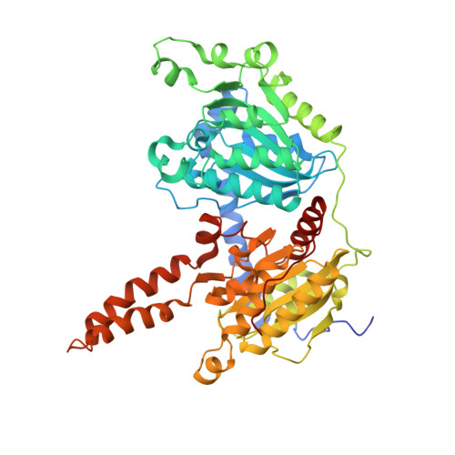
    3-Methylcrotonyl-CoA carboxylase (MCC), a member of the biotin-dependent carboxylase superfamily, is essential for the metabolism of leucine, and deficient mutations in this enzyme are linked to methylcrotonylglycinuria (MCG) and other serious diseases in humans. MCC has strong sequence conservation with propionyl-CoA carboxylase (PCC), and their holoenzymes are both 750-kilodalton (kDa) α(6)β(6) dodecamers. Therefore the architecture of the MCC holoenzyme is expected to be highly similar to that of PCC. Here we report the crystal structures of the Pseudomonas aeruginosa MCC (PaMCC) holoenzyme, alone and in complex with coenzyme A. Surprisingly, the structures show that the architecture and overall shape of PaMCC are markedly different when compared to PCC. The α-subunits show trimeric association in the PaMCC holoenzyme, whereas they have no contacts with each other in PCC. Moreover, the positions of the two domains in the β-subunit of PaMCC are swapped relative to those in PCC. This structural information establishes a foundation for understanding the disease-causing mutations of MCC and provides new insights into the catalytic mechanism and evolution of biotin-dependent carboxylases. The large structural differences between MCC and PCC also have general implications for the relationship between sequence conservation and structural similarity.


  • Organizational Affiliation

    Department of Biological Sciences, Columbia University, New York, New York 10027, USA.


Macromolecules
Find similar proteins by:  (by identity cutoff)  |  3D Structure
Entity ID: 1
MoleculeChains Sequence LengthOrganismDetailsImage
Methylcrotonyl-CoA carboxylase, alpha-subunit
A, C, E, G, I
A, C, E, G, I, K
655Pseudomonas aeruginosaMutation(s): 0 
Gene Names: liuDPA2012
EC: 6.4.1.4
UniProt
Find proteins for Q9I299 (Pseudomonas aeruginosa (strain ATCC 15692 / DSM 22644 / CIP 104116 / JCM 14847 / LMG 12228 / 1C / PRS 101 / PAO1))
Explore Q9I299 
Go to UniProtKB:  Q9I299
Entity Groups  
Sequence Clusters30% Identity50% Identity70% Identity90% Identity95% Identity100% Identity
UniProt GroupQ9I299
Sequence Annotations
Expand
  • Reference Sequence
Find similar proteins by:  (by identity cutoff)  |  3D Structure
Entity ID: 2
MoleculeChains Sequence LengthOrganismDetailsImage
Methylcrotonyl-CoA carboxylase, beta-subunit
B, D, F, H, J
B, D, F, H, J, L
555Pseudomonas aeruginosaMutation(s): 0 
Gene Names: liuBPA2014
EC: 6.4.1.4
UniProt
Find proteins for Q9I297 (Pseudomonas aeruginosa (strain ATCC 15692 / DSM 22644 / CIP 104116 / JCM 14847 / LMG 12228 / 1C / PRS 101 / PAO1))
Explore Q9I297 
Go to UniProtKB:  Q9I297
Entity Groups  
Sequence Clusters30% Identity50% Identity70% Identity90% Identity95% Identity100% Identity
UniProt GroupQ9I297
Sequence Annotations
Expand
  • Reference Sequence
Experimental Data & Validation

Experimental Data

  • Method: X-RAY DIFFRACTION
  • Resolution: 3.50 Å
  • R-Value Free: 0.292 
  • R-Value Work: 0.234 
  • R-Value Observed: 0.234 
  • Space Group: P 1 21 1
Unit Cell:
Length ( Å )Angle ( ˚ )
a = 131.51α = 90
b = 255.34β = 95.74
c = 152.67γ = 90
Software Package:
Software NamePurpose
HKL-2000data collection
PHASERphasing
CNSrefinement
DENZOdata reduction
SCALEPACKdata scaling

Structure Validation

View Full Validation Report


Ligand Structure Quality Assessment 


Entry History 

Deposition Data

Revision History  (Full details and data files)

  • Version 1.0: 2011-12-14
    Type: Initial release
  • Version 1.1: 2013-01-23
    Changes: Database references, Structure summary