


The crystal structure of Mandelate racemase/muconate lactonizing enzyme from Roseiflexus sp.
Zhang, Z., Almo, S.C., Swaminathan, S.To be published.
Experimental Data Snapshot
Starting Model: experimental
View more details
wwPDB Validation 3D Report Full Report

Entity ID: 1 | |||||
|---|---|---|---|---|---|
| Molecule | Chains | Sequence Length | Organism | Details | Image |
| Mandelate racemase/muconate lactonizing enzyme, C-terminal domain protein | 393 | Roseiflexus sp. RS-1 | Mutation(s): 0 Gene Names: RoseRS_2982 | 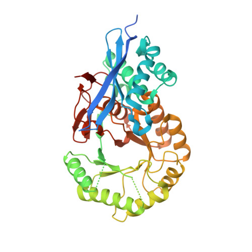 | |
Entity Groups | |||||
| Sequence Clusters | 30% Identity50% Identity70% Identity90% Identity95% Identity100% Identity | ||||
Sequence AnnotationsExpand | |||||
Reference Sequence | |||||
| Ligands 1 Unique | |||||
|---|---|---|---|---|---|
| ID | Chains | Name / Formula / InChI Key | 2D Diagram | 3D Interactions | |
| SO4 Download:Ideal Coordinates CCD File | C [auth A], D [auth B] | SULFATE ION O4 S QAOWNCQODCNURD-UHFFFAOYSA-L |  | ||
| Length ( Å ) | Angle ( ˚ ) |
|---|---|
| a = 209.888 | α = 90 |
| b = 209.888 | β = 90 |
| c = 116.601 | γ = 120 |
| Software Name | Purpose |
|---|---|
| CBASS | data collection |
| PHASER | phasing |
| PHENIX | refinement |
| HKL-2000 | data reduction |
| HKL-2000 | data scaling |


