3U9G

Crystal structure of the Zinc finger antiviral protein


Experimental Data Snapshot

  • Method: X-RAY DIFFRACTION
  • Resolution: 1.80 Å
  • R-Value Free: 0.228 
  • R-Value Work: 0.191 
  • R-Value Observed: 0.193 

wwPDB Validation   3D Report Full Report


This is version 1.2 of the entry. See complete history


Literature

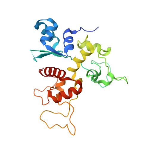
Structure of N-terminal domain of ZAP indicates how a zinc-finger protein recognizes complex RNA.

Chen, S.Xu, Y.Zhang, K.Wang, X.Sun, J.Gao, G.Liu, Y.

(2012) Nat Struct Mol Biol 19: 430-435

  • DOI: https://doi.org/10.1038/nsmb.2243
  • Primary Citation of Related Structures:  
    3U9G

  • PubMed Abstract: 

    Zinc-finger antiviral protein (ZAP) is a host factor that specifically inhibits the replication of certain viruses, such as HIV-1, by targeting viral mRNA for degradation. How ZAP recognizes its target RNA has been unclear. Here we report the crystal structure of the N-terminal domain of rat ZAP (NZAP225), the major functional domain. The overall structure of NZAP225 resembles a tractor, with four zinc-finger motifs located at the bottom. Structural and functional analyses identified multiple positively charged residues and two putative RNA-binding cavities forming a large putative RNA-binding cleft. ZAP molecules interact to form a dimer that binds to a ZAP-responsive RNA molecule containing two ZAP-binding modules. These results provide insights into how ZAP binds specifically to complex target RNA.


  • Organizational Affiliation

    State Key Laboratory of Biomacromolecules, Institute of Biophysics, Chinese Academy of Sciences, Beijing, China.


Macromolecules
Find similar proteins by:  (by identity cutoff)  |  3D Structure
Entity ID: 1
MoleculeChains Sequence LengthOrganismDetailsImage
Zinc finger CCCH-type antiviral protein 1229Rattus norvegicusMutation(s): 0 
Gene Names: Zc3hav1Zap
UniProt
Find proteins for Q8K3Y6 (Rattus norvegicus)
Explore Q8K3Y6 
Go to UniProtKB:  Q8K3Y6
Entity Groups  
Sequence Clusters30% Identity50% Identity70% Identity90% Identity95% Identity100% Identity
UniProt GroupQ8K3Y6
Sequence Annotations
Expand
  • Reference Sequence
Experimental Data & Validation

Experimental Data

  • Method: X-RAY DIFFRACTION
  • Resolution: 1.80 Å
  • R-Value Free: 0.228 
  • R-Value Work: 0.191 
  • R-Value Observed: 0.193 
  • Space Group: P 32 2 1
Unit Cell:
Length ( Å )Angle ( ˚ )
a = 52.894α = 90
b = 52.894β = 90
c = 138.275γ = 120
Software Package:
Software NamePurpose
MAR345dtbdata collection
SHELXSphasing
PHENIXrefinement
HKL-2000data reduction
HKL-2000data scaling

Structure Validation

View Full Validation Report


Entry History 

Deposition Data

Revision History  (Full details and data files)

  • Version 1.0: 2012-03-14
    Type: Initial release
  • Version 1.1: 2013-07-17
    Changes: Database references
  • Version 1.2: 2024-03-20
    Changes: Data collection, Database references, Derived calculations