Low-density crystal packing of human protein kinase CK2 catalytic subunit in complex with resorufin or other ligands: a tool to study the unique hinge-region plasticity of the enzyme without packing bias.
Klopffleisch, K., Issinger, O.G., Niefind, K.(2012) Acta Crystallogr D Biol Crystallogr 68: 883-892
- PubMed: 22868753 Search on PubMed
- DOI: https://doi.org/10.1107/S0907444912016587
- Primary Citation Related Structures:
3U87, 3U9C - PubMed Abstract:
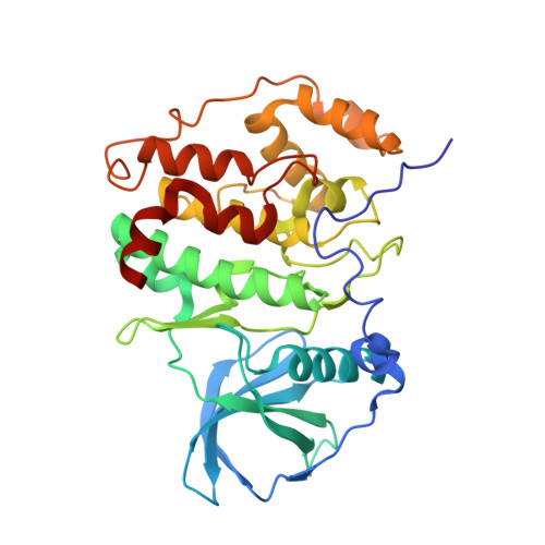
A low-resolution structure of the catalytic subunit CK2α of human protein kinase CK2 (formerly known as casein kinase 2) in complex with the ATP-competitive inhibitor resorufin is presented. The structure supplements previous human CK2α structures in which the interdomain hinge/helix αD region adopts a closed conformation correlating to a canonically established catalytic spine as is typical for eukaryotic protein kinases. In the corresponding crystal packing the hinge/helix αD region is nearly unaffected by crystal contacts, so that largely unbiased conformational adaptions are possible. This is documented by published human CK2α structures with the same crystal packing but with an open hinge/helix αD region, one of which has been redetermined here with a higher symmetry. An overview of all published human CK2α crystal packings serves as the basis for a discussion of the factors that determine whether the open or the closed hinge/helix αD conformation is adopted. Lyotropic salts in crystallization support the closed conformation, in which the Phe121 side chain complements the hydrophobic catalytic spine ensemble. Consequently, genuine ligand effects on the hinge/helix αD conformation can be best studied under moderate salt conditions. Ligands that stabilize either the open or the closed conformation by hydrogen bonds are known, but a general rule is not yet apparent.
- Department für Chemie, Institut für Biochemie, Universität zu Köln, Otto-Fischer-Strasse 12-14, D-50674 Köln, Germany.
Organizational Affiliation: