Structural analysis of smooth muscle tropomyosin alpha and beta isoforms.
Rao, J.N., Rivera-Santiago, R., Li, X.E., Lehman, W., Dominguez, R.(2012) J Biol Chem 287: 3165-3174
- PubMed: 22119916 Search on PubMedSearch on PubMed Central
- DOI: https://doi.org/10.1074/jbc.M111.307330
- Primary Citation Related Structures:
3U1A, 3U1C, 3U59 - PubMed Abstract:
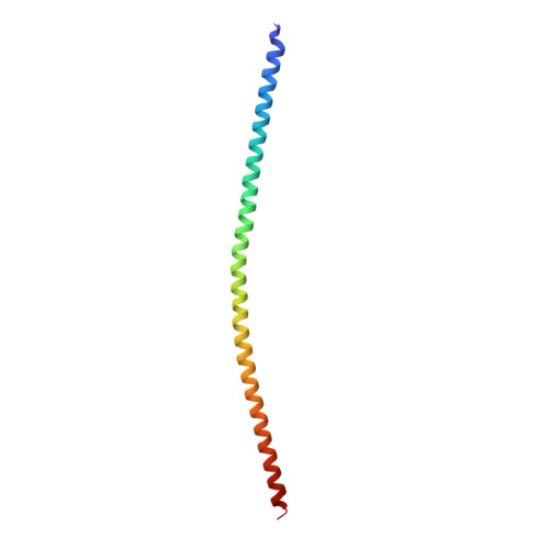
A large number of tropomyosin (Tm) isoforms function as gatekeepers of the actin filament, controlling the spatiotemporal access of actin-binding proteins to specialized actin networks. Residues ∼40-80 vary significantly among Tm isoforms, but the impact of sequence variation on Tm structure and interactions with actin is poorly understood, because structural studies have focused on skeletal muscle Tmα. We describe structures of N-terminal fragments of smooth muscle Tmα and Tmβ (sm-Tmα and sm-Tmβ). The 2.0-Å structure of sm-Tmα81 (81-aa) resembles that of skeletal Tmα, displaying a similar super-helical twist matching the contours of the actin filament. The 1.8-Å structure of sm-Tmα98 (98-aa) unexpectedly reveals an antiparallel coiled coil, with the two chains staggered by only 4 amino acids and displaying hydrophobic core interactions similar to those of the parallel dimer. In contrast, the 2.5-Å structure of sm-Tmβ98, containing Gly-Ala-Ser at the N terminus to mimic acetylation, reveals a parallel coiled coil. None of the structures contains coiled-coil stabilizing elements, favoring the formation of head-to-tail overlap complexes in four of five crystallographically independent parallel dimers. These complexes show similarly arranged 4-helix bundles stabilized by hydrophobic interactions, but the extent of the overlap varies between sm-Tmβ98 and sm-Tmα81 from 2 to 3 helical turns. The formation of overlap complexes thus appears to be an intrinsic property of the Tm coiled coil, with the specific nature of hydrophobic contacts determining the extent of the overlap. Overall, the results suggest that sequence variation among Tm isoforms has a limited effect on actin binding but could determine its gatekeeper function.
- Department of Physiology, Perelman School of Medicine, University of Pennsylvania, Philadelphia, Pennsylvania 19104, USA.
Organizational Affiliation: