3U53

Crystal structure of human Ap4A hydrolase


Experimental Data Snapshot

  • Method: X-RAY DIFFRACTION
  • Resolution: 2.71 Å
  • R-Value Free: 0.284 
  • R-Value Work: 0.213 
  • R-Value Observed: 0.217 

wwPDB Validation   3D Report Full Report


This is version 1.2 of the entry. See complete history


Literature

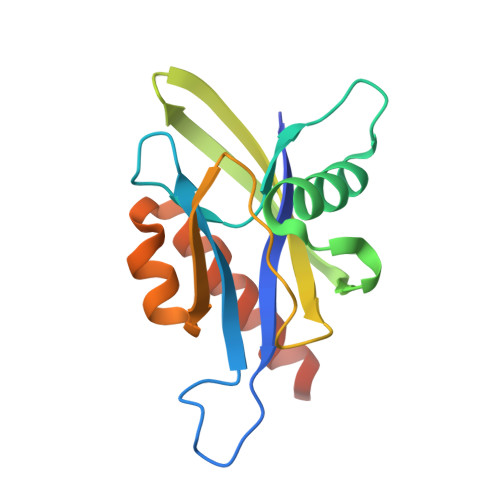
Crystal structure of wild-type and mutant human Ap4A hydrolase

Ge, H.H.Chen, X.F.Yang, W.L.Niu, L.W.Teng, M.K.

(2013) Biochem Biophys Res Commun 432: 16-21

  • DOI: https://doi.org/10.1016/j.bbrc.2013.01.095
  • Primary Citation of Related Structures:  
    3U53, 4ICK, 4IJX

  • PubMed Abstract: 

    Ap4A hydrolase (asymmetrical diadenosine tetraphosphate hydrolase, EC 3.6.1.17), an enzyme involved in a number of biological processes, is characterized as cleaving the polyphosphate chain at the fourth phosphate from the bound adenosine moiety. This paper presents the crystal structure of wild-type and E58A mutant human Ap4A hydrolase. Similar to the canonical Nudix fold, human Ap4A hydrolase shows the common αβα-sandwich architecture. Interestingly, two sulfate ions and one diphosphate coordinated with some conserved residues were observed in the active cleft, which affords a better understanding of a possible mode of substrate binding.


  • Organizational Affiliation

    Institute of Health Sciences and Modern Experiment Technology Center, Anhui University, Hefei 230601, People's Republic of China. hhge@ahu.edu.cn


Macromolecules
Find similar proteins by:  (by identity cutoff)  |  3D Structure
Entity ID: 1
MoleculeChains Sequence LengthOrganismDetailsImage
Bis(5'-nucleosyl)-tetraphosphatase [asymmetrical]
A, B, C, D
155Homo sapiensMutation(s): 0 
Gene Names: NUDT2
EC: 3.6.1.17
UniProt & NIH Common Fund Data Resources
Find proteins for P50583 (Homo sapiens)
Explore P50583 
Go to UniProtKB:  P50583
PHAROS:  P50583
GTEx:  ENSG00000164978 
Entity Groups  
Sequence Clusters30% Identity50% Identity70% Identity90% Identity95% Identity100% Identity
UniProt GroupP50583
Sequence Annotations
Expand
  • Reference Sequence
Small Molecules
Ligands 2 Unique
IDChains Name / Formula / InChI Key2D Diagram3D Interactions
SO4
Query on SO4

Download Ideal Coordinates CCD File 
E [auth A]
F [auth A]
G [auth A]
I [auth B]
J [auth B]
E [auth A],
F [auth A],
G [auth A],
I [auth B],
J [auth B],
K [auth B],
L [auth B],
M [auth C],
N [auth C],
O [auth C],
P [auth C],
Q [auth C],
R [auth D],
S [auth D],
T [auth D],
U [auth D],
V [auth D]
SULFATE ION
O4 S
QAOWNCQODCNURD-UHFFFAOYSA-L
GOL
Query on GOL

Download Ideal Coordinates CCD File 
H [auth A]GLYCEROL
C3 H8 O3
PEDCQBHIVMGVHV-UHFFFAOYSA-N
Experimental Data & Validation

Experimental Data

  • Method: X-RAY DIFFRACTION
  • Resolution: 2.71 Å
  • R-Value Free: 0.284 
  • R-Value Work: 0.213 
  • R-Value Observed: 0.217 
  • Space Group: P 43
Unit Cell:
Length ( Å )Angle ( ˚ )
a = 72.485α = 90
b = 72.485β = 90
c = 133.491γ = 90
Software Package:
Software NamePurpose
HKL-2000data collection
PHASERphasing
REFMACrefinement
HKL-2000data reduction
HKL-2000data scaling

Structure Validation

View Full Validation Report


Entry History 

Deposition Data

Revision History  (Full details and data files)

  • Version 1.0: 2012-10-10
    Type: Initial release
  • Version 1.1: 2013-03-20
    Changes: Database references
  • Version 1.2: 2023-11-01
    Changes: Data collection, Database references, Derived calculations, Refinement description