3U52 | pdb_00003u52

X-ray Crystal Structure of Xenon-Pressurized Phenol Hydroxylase from Pseudomonas sp. OX1


Experimental Data Snapshot

  • Method: X-RAY DIFFRACTION
  • Resolution: 1.95 Å
  • R-Value Free: 
    0.228 (Depositor), 0.185 (DCC) 
  • R-Value Work: 
    0.183 (Depositor) 
  • R-Value Observed: 
    0.185 (Depositor) 

Starting Model: experimental
View more details

wwPDB Validation 3D Report Full Report

Validation slider image for 3U52

Ligand Structure Quality Assessment 


This is version 1.2 of the entry. See complete history

Literature

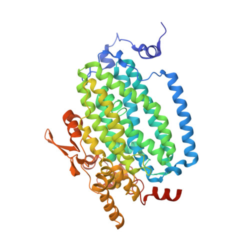
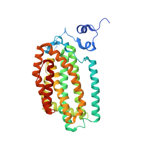
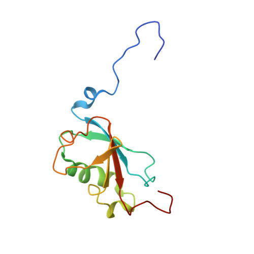
Analysis of Substrate Access to Active Sites in Bacterial Multicomponent Monooxygenase Hydroxylases: X-ray Crystal Structure of Xenon-Pressurized Phenol Hydroxylase from Pseudomonas sp. OX1.

McCormick, M.S.Lippard, S.J.

(2011) Biochemistry 50: 11058-11069

  • DOI: https://doi.org/10.1021/bi201248b
  • Primary Citation Related Structures: 
    3U52

  • PubMed Abstract: 

    In all structurally characterized bacterial multicomponent monooxygenase (BMM) hydroxylase proteins, a series of hydrophobic cavities in the α-subunit trace a conserved path from the protein exterior to the carboxylate-bridged diiron active site. This study examines these cavities as a potential route for transport of dioxygen to the active site by crystallographic characterization of a xenon-pressurized sample of the hydroxylase component of phenol hydroxylase from Pseudomonas sp. OX1. Computational analyses of the hydrophobic cavities in the hydroxylase α-subunits of phenol hydroxylase (PHH), soluble methane monooxygenase (MMOH), and toluene/o-xylene monooxygenase (ToMOH) are also presented. The results, together with previous findings from crystallographic studies of xenon-pressurized sMMO hydroxylase, clearly identify the propensity for these cavities to bind hydrophobic gas molecules in the protein interior. This proposed functional role is supported by recent stopped flow kinetic studies of ToMOH variants [Song, W. J., et al. (2011) Proc. Natl. Acad. Sci. U.S.A.108, 14795-14800]. In addition to information about the Xe sites, the structure determination revealed significantly weakened binding of regulatory protein to the hydroxylase in comparison to that in the previously reported structure of PHH, as well as the presence of a newly identified metal-binding site in the α-subunit that adopts a linear coordination environment consistent with Cu(I), and a glycerol molecule bound to Fe1 in a fashion that is unique among hydrocarbon-diiron site adducts reported to date in BMM hydroxylase structures. Finally, a comparative analysis of the α-subunit structures of PHH, MMOH, and ToMOH details proposed routes for the other three BMM substrates, the hydrocarbon, electrons, and protons, comprising cavities, channels, hydrogen-bonding networks, and pores in the structures of their α-subunits.


  • Organizational Affiliation
    • Department of Chemistry, Massachusetts Institute of Technology, Cambridge, Massachusetts 02139, United States.

Macromolecule Content 

  • Total Structure Weight: 228.85 kDa 
  • Atom Count: 16,039 
  • Modeled Residue Count: 1,869 
  • Deposited Residue Count: 1,926 
  • Unique protein chains: 3

Macromolecules

Find similar proteins by:|  3D Structure
Entity ID: 1
MoleculeChains  Sequence LengthOrganismDetailsImage
Phenol hydroxylase component phN
A, B
511Stutzerimonas stutzeriMutation(s): 0 
Gene Names: phN
UniProt
Find proteins for Q84AQ2 (Stutzerimonas stutzeri)
Explore Q84AQ2 
Go to UniProtKB:  Q84AQ2
Entity Groups
Sequence Clusters30% Identity50% Identity70% Identity90% Identity95% Identity100% Identity
UniProt GroupQ84AQ2
Sequence Annotations
Expand
Reference Sequence
Find similar proteins by:|  3D Structure
Entity ID: 2
MoleculeChains  Sequence LengthOrganismDetailsImage
Phenol hydroxylase component phL
C, D
333Stutzerimonas stutzeriMutation(s): 0 
Gene Names: phL
UniProt
Find proteins for Q84AQ4 (Stutzerimonas stutzeri)
Explore Q84AQ4 
Go to UniProtKB:  Q84AQ4
Entity Groups
Sequence Clusters30% Identity50% Identity70% Identity90% Identity95% Identity100% Identity
UniProt GroupQ84AQ4
Sequence Annotations
Expand
Reference Sequence
Find similar proteins by:|  3D Structure
Entity ID: 3
MoleculeChains  Sequence LengthOrganismDetailsImage
Phenol hydroxylase component phO
E, F
119Stutzerimonas stutzeriMutation(s): 0 
Gene Names: phO
UniProt
Find proteins for Q84AQ1 (Stutzerimonas stutzeri)
Explore Q84AQ1 
Go to UniProtKB:  Q84AQ1
Entity Groups
Sequence Clusters30% Identity50% Identity70% Identity90% Identity95% Identity100% Identity
UniProt GroupQ84AQ1
Sequence Annotations
Expand
Reference Sequence

Small Molecules

Ligands 7 Unique
IDChains Name / Formula / InChI Key2D Diagram3D Interactions
EPE

Query on EPE


Download:Ideal Coordinates CCD File
EA [auth B]4-(2-HYDROXYETHYL)-1-PIPERAZINE ETHANESULFONIC ACID
C8 H18 N2 O4 S
JKMHFZQWWAIEOD-UHFFFAOYSA-N
MPO

Query on MPO


Download:Ideal Coordinates CCD File
DA [auth B]3[N-MORPHOLINO]PROPANE SULFONIC ACID
C7 H15 N O4 S
DVLFYONBTKHTER-UHFFFAOYSA-N
XE

Query on XE


Download:Ideal Coordinates CCD File
HA [auth B]
IA [auth B]
JA [auth B]
KA [auth B]
LA [auth B]
HA [auth B],
IA [auth B],
JA [auth B],
KA [auth B],
LA [auth B],
MA [auth B],
NA [auth B],
O [auth A],
OA [auth B],
P [auth A],
PA [auth B],
Q [auth A],
QA [auth B],
R [auth A],
RA [auth B],
S [auth A],
T [auth A],
U [auth A],
UA [auth C],
V [auth A],
W [auth A],
WA [auth D],
X [auth A],
Y [auth A]
XENON
Xe
FHNFHKCVQCLJFQ-UHFFFAOYSA-N
GOL

Query on GOL


Download:Ideal Coordinates CCD File
FA [auth B]
GA [auth B]
K [auth A]
L [auth A]
M [auth A]
FA [auth B],
GA [auth B],
K [auth A],
L [auth A],
M [auth A],
N [auth A],
SA [auth C],
TA [auth C],
VA [auth D],
XA [auth F]
GLYCEROL
C3 H8 O3
PEDCQBHIVMGVHV-UHFFFAOYSA-N
ZN

Query on ZN


Download:Ideal Coordinates CCD File
BA [auth B],
I [auth A]
ZINC ION
Zn
PTFCDOFLOPIGGS-UHFFFAOYSA-N
CU

Query on CU


Download:Ideal Coordinates CCD File
CA [auth B],
J [auth A]
COPPER (II) ION
Cu
JPVYNHNXODAKFH-UHFFFAOYSA-N
FE

Query on FE


Download:Ideal Coordinates CCD File
AA [auth B],
G [auth A],
H [auth A],
Z [auth B]
FE (III) ION
Fe
VTLYFUHAOXGGBS-UHFFFAOYSA-N

Experimental Data & Validation

Experimental Data

  • Method: X-RAY DIFFRACTION
  • Resolution: 1.95 Å
  • R-Value Free:  0.228 (Depositor), 0.185 (DCC) 
  • R-Value Work:  0.183 (Depositor) 
  • R-Value Observed: 0.185 (Depositor) 
Space Group: P 21 21 21
Unit Cell:
Length ( Å )Angle ( ˚ )
a = 83.942α = 90
b = 141.761β = 90
c = 181.207γ = 90
Software Package:
Software NamePurpose
Blu-Icedata collection
EPMRphasing
REFMACrefinement
HKL-2000data reduction
HKL-2000data scaling

Structure Validation

View Full Validation Report


Ligand Structure Quality Assessment 


Entry History 

Deposition Data

Revision History  (Full details and data files)

  • Version 1.0: 2011-12-14
    Type: Initial release
  • Version 1.1: 2012-01-11
    Changes: Database references
  • Version 1.2: 2023-09-13
    Changes: Data collection, Database references, Derived calculations, Refinement description