3U3D

Plasmodium falciparum Sir2A preferentially hydrolyzes medium and long chain fatty acyl lysine


Experimental Data Snapshot

  • Method: X-RAY DIFFRACTION
  • Resolution: 2.40 Å
  • R-Value Free: 0.276 
  • R-Value Work: 0.224 
  • R-Value Observed: 0.226 

wwPDB Validation   3D Report Full Report


This is version 1.2 of the entry. See complete history


Literature

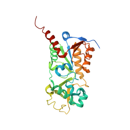
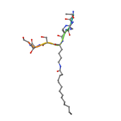
Plasmodium falciparum Sir2A Preferentially Hydrolyzes Medium and Long Chain Fatty Acyl Lysine

Zhu, A.Y.Zhou, Y.Khan, S.Deitsch, K.W.Hao, Q.Lin, H.

(2011) ACS Chem Biol 

  • DOI: https://doi.org/10.1021/cb200230x
  • Primary Citation of Related Structures:  
    3U31, 3U3D

  • PubMed Abstract: 

    Plasmodium falciparum Sir2A (PfSir2A), a member of the sirtuin family of nicotinamide adenine dinucleotide-dependent deacetylases, has been shown to regulate the expression of surface antigens to evade the detection by host immune surveillance. It is thought that PfSir2A achieves this by deacetylating histones. However, the deacetylase activity of PfSir2A is weak. Here we present enzymology and structural evidence supporting that PfSir2A catalyzes the hydrolysis of medium and long chain fatty acyl groups from lysine residues more efficiently. Furthermore, P. falciparum proteins are found to contain such fatty acyl lysine modifications that can be removed by purified PfSir2A in vitro. Together, the data suggest that the physiological function of PfSir2A in antigen variation may be achieved by removing medium and long chain fatty acyl groups from protein lysine residues. The robust activity of PfSir2A would also facilitate the development of PfSir2A inhibitors, which may have therapeutic value in malaria treatment.


Macromolecules
Find similar proteins by:  (by identity cutoff)  |  3D Structure
Entity ID: 1
MoleculeChains Sequence LengthOrganismDetailsImage
Transcriptional regulatory protein sir2 homologue290Plasmodium falciparum 3D7Mutation(s): 0 
Gene Names: PF13_0152Sir2
EC: 3.5.1.17
UniProt
Find proteins for Q8IE47 (Plasmodium falciparum (isolate 3D7))
Explore Q8IE47 
Go to UniProtKB:  Q8IE47
Entity Groups  
Sequence Clusters30% Identity50% Identity70% Identity90% Identity95% Identity100% Identity
UniProt GroupQ8IE47
Sequence Annotations
Expand
  • Reference Sequence

Find similar proteins by:  Sequence   |   3D Structure  

Entity ID: 2
MoleculeChains Sequence LengthOrganismDetailsImage
histone 3 myristoyl lysine 9 peptide12N/AMutation(s): 0 
UniProt & NIH Common Fund Data Resources
Find proteins for P68431 (Homo sapiens)
Explore P68431 
Go to UniProtKB:  P68431
PHAROS:  P68431
Entity Groups  
Sequence Clusters30% Identity50% Identity70% Identity90% Identity95% Identity100% Identity
UniProt GroupP68431
Sequence Annotations
Expand
  • Reference Sequence
Small Molecules
Modified Residues  1 Unique
IDChains TypeFormula2D DiagramParent
MYK
Query on MYK
B
L-PEPTIDE LINKINGC20 H40 N2 O3LYS
Experimental Data & Validation

Experimental Data

  • Method: X-RAY DIFFRACTION
  • Resolution: 2.40 Å
  • R-Value Free: 0.276 
  • R-Value Work: 0.224 
  • R-Value Observed: 0.226 
  • Space Group: P 21 21 2
Unit Cell:
Length ( Å )Angle ( ˚ )
a = 32.511α = 90
b = 103.328β = 90
c = 105.512γ = 90
Software Package:
Software NamePurpose
DENZOdata reduction
SCALEPACKdata scaling
REFMACrefinement
PDB_EXTRACTdata extraction
CrystalCleardata collection
HKL-2000data reduction
MOLREPphasing

Structure Validation

View Full Validation Report


Entry History 

Deposition Data

Revision History  (Full details and data files)

  • Version 1.0: 2011-11-09
    Type: Initial release
  • Version 1.1: 2017-11-08
    Changes: Refinement description
  • Version 1.2: 2023-11-01
    Changes: Data collection, Database references, Derived calculations, Refinement description