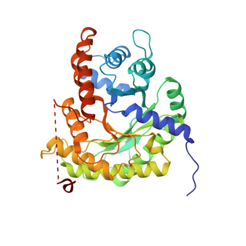
3TU9 | pdb_00003tu9

Crystal structure of rabbit muscle aldolase bound with 5-O-methyl mannitol 1,6-phosphate


Experimental Data Snapshot

  • Method: X-RAY DIFFRACTION
  • Resolution: 2.09 Å
  • R-Value Free: 
    0.201 (Depositor), 0.200 (DCC) 
  • R-Value Work: 
    0.156 (Depositor), 0.150 (DCC) 
  • R-Value Observed: 
    0.160 (Depositor) 

Starting Model: experimental
View more details

wwPDB Validation   3D Report Full Report


Ligand Structure Quality Assessment 

Created with Raphaël 2.3.0Worse 01 BetterLigand structure goodness of fit to experimental dataBest fitted 5MMClick on this verticalbar to view details

This is version 1.2 of the entry. See complete history


Literature

Mannitol Bis-phosphate Based Inhibitors of Fructose 1,6-Bisphosphate Aldolases.

Mabiala-Bassiloua, C.G.Arthus-Cartier, G.Hannaert, V.Therisod, H.Sygusch, J.Therisod, M.

(2011) ACS Med Chem Lett 2: 804-808

  • DOI: https://doi.org/10.1021/ml200129s
  • Primary Citation of Related Structures:  
    3TU9

  • PubMed Abstract: 

    Several 5-O-alkyl- and 5-C-alkyl-mannitol bis-phosphates were synthesized and comparatively assayed as inhibitors of fructose bis-phosphate aldolases (Fbas) from rabbit muscle (taken as surrogate model of the human enzyme) and from Trypanosoma brucei. A limited selectivity was found in several instances. Crystallographic studies confirm that the 5-O-methyl derivative binds competitively with substrate and the 5-O-methyl moiety penetrating deeper into a shallow hydrophobic pocket at the active site. This observation can lead to the preparation of selective competitive or irreversible inhibitors of the parasite Fba.


  • Organizational Affiliation

    ECBB, ICMMO (UMR 8182), LabEx LERMIT, Université Paris-Sud , UMR 8182, F-91405 Orsay, France.


Macromolecules
Find similar proteins by:  (by identity cutoff)  |  3D Structure
Entity ID: 1
MoleculeChains Sequence LengthOrganismDetailsImage
Fructose-bisphosphate aldolase A
A, B, C, D
363Oryctolagus cuniculusMutation(s): 0 
Gene Names: ALDOA
EC: 4.1.2.13
UniProt
Find proteins for P00883 (Oryctolagus cuniculus)
Explore P00883 
Go to UniProtKB:  P00883
Entity Groups  
Sequence Clusters30% Identity50% Identity70% Identity90% Identity95% Identity100% Identity
UniProt GroupP00883
Sequence Annotations
Expand
  • Reference Sequence
Experimental Data & Validation

Experimental Data

  • Method: X-RAY DIFFRACTION
  • Resolution: 2.09 Å
  • R-Value Free:  0.201 (Depositor), 0.200 (DCC) 
  • R-Value Work:  0.156 (Depositor), 0.150 (DCC) 
  • R-Value Observed: 0.160 (Depositor) 
Space Group: P 1 21 1
Unit Cell:
Length ( Å )Angle ( ˚ )
a = 82.669α = 90
b = 103.056β = 98.34
c = 84.458γ = 90
Software Package:
Software NamePurpose
HKL-2000data collection
PHENIXmodel building
PHENIXrefinement
HKL-2000data reduction
HKL-2000data scaling
PHENIXphasing

Structure Validation

View Full Validation Report


Ligand Structure Quality Assessment 

Created with Raphaël 2.3.0Worse 01 BetterLigand structure goodness of fit to experimental dataBest fitted 5MMClick on this verticalbar to view details

Entry History 

Deposition Data

Revision History  (Full details and data files)

  • Version 1.0: 2011-10-12
    Type: Initial release
  • Version 1.1: 2018-04-18
    Changes: Data collection, Database references
  • Version 1.2: 2023-09-13
    Changes: Data collection, Database references, Derived calculations, Refinement description