3TTE | pdb_00003tte

Crystal structure of enolase brado_4202 (target EFI-501651) from Bradyrhizobium complexed with magnesium and mandelic acid


Experimental Data Snapshot

  • Method: X-RAY DIFFRACTION
  • Resolution: 2.00 Å
  • R-Value Free: 
    0.263 (Depositor), 0.259 (DCC) 
  • R-Value Work: 
    0.204 (Depositor), 0.204 (DCC) 
  • R-Value Observed: 
    0.206 (Depositor) 

Starting Model: experimental
View more details

wwPDB Validation 3D Report Full Report

Validation slider image for 3TTE

Ligand Structure Quality Assessment 


This is version 1.2 of the entry. See complete history

Literature

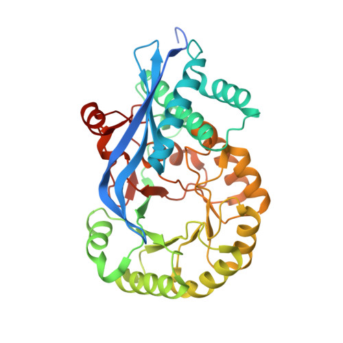
Crystal Structure of Mandelate Racemase from Bradyrhizobium Sp. Ors278

Patskovsky, Y.Kim, J.Toro, R.Bhosle, R.Hillerich, B.Seidel, R.D.Washington, E.Scott Glenn, A.Chowdhury, S.Evans, B.Hammond, J.Zencheck, W.D.Imker, H.J.Gerlt, J.A.Almo, S.C.

To be published.

Macromolecule Content 

  • Total Structure Weight: 82.85 kDa 
  • Atom Count: 5,680 
  • Modeled Residue Count: 720 
  • Deposited Residue Count: 766 
  • Unique protein chains: 1

Macromolecules

Find similar proteins by:|  3D Structure
Entity ID: 1
MoleculeChains  Sequence LengthOrganismDetailsImage
Mandelate racemase/muconate lactonizing enzyme
A, B
383Bradyrhizobium sp. ORS 278Mutation(s): 0 
Gene Names: BRADO4202
EC: 5.1.2.2 (PDB Primary Data), 5.5.1.1 (PDB Primary Data)
UniProt
Find proteins for A0ACD6B9G9 (Bradyrhizobium sp. (strain ORS 278))
Explore A0ACD6B9G9 
Go to UniProtKB:  A0ACD6B9G9
Entity Groups
Sequence Clusters30% Identity50% Identity70% Identity90% Identity95% Identity100% Identity
UniProt GroupA0ACD6B9G9
Sequence Annotations
Expand
Reference Sequence

Small Molecules

Ligands 4 Unique
IDChains Name / Formula / InChI Key2D Diagram3D Interactions
SMN

Query on SMN


Download:Ideal Coordinates CCD File
D [auth A](S)-MANDELIC ACID
C8 H8 O3
IWYDHOAUDWTVEP-ZETCQYMHSA-N
GOL

Query on GOL


Download:Ideal Coordinates CCD File
E [auth A],
F [auth A],
H [auth B]
GLYCEROL
C3 H8 O3
PEDCQBHIVMGVHV-UHFFFAOYSA-N
FMT

Query on FMT


Download:Ideal Coordinates CCD File
I [auth B]FORMIC ACID
C H2 O2
BDAGIHXWWSANSR-UHFFFAOYSA-N
MG

Query on MG


Download:Ideal Coordinates CCD File
C [auth A],
G [auth B]
MAGNESIUM ION
Mg
JLVVSXFLKOJNIY-UHFFFAOYSA-N

Experimental Data & Validation

Experimental Data

  • Method: X-RAY DIFFRACTION
  • Resolution: 2.00 Å
  • R-Value Free:  0.263 (Depositor), 0.259 (DCC) 
  • R-Value Work:  0.204 (Depositor), 0.204 (DCC) 
  • R-Value Observed: 0.206 (Depositor) 
Space Group: I 2 2 2
Unit Cell:
Length ( Å )Angle ( ˚ )
a = 109.949α = 90
b = 118.867β = 90
c = 130.571γ = 90
Software Package:
Software NamePurpose
PHASERphasing
REFMACrefinement
HKL-3000data reduction
HKL-3000data scaling

Structure Validation

View Full Validation Report


Ligand Structure Quality Assessment 


Entry History 

Revision History  (Full details and data files)

  • Version 1.0: 2011-09-28
    Type: Initial release
  • Version 1.1: 2012-02-22
    Changes: Database references, Structure summary
  • Version 1.2: 2023-09-13
    Changes: Data collection, Database references, Derived calculations, Refinement description