3TM9

Y29A mutant of Vitreoscilla stercoraria hemoglobin


Experimental Data Snapshot

  • Method: X-RAY DIFFRACTION
  • Resolution: 1.72 Å
  • R-Value Free: 0.206 
  • R-Value Work: 0.172 
  • R-Value Observed: 0.176 

wwPDB Validation   3D Report Full Report


Ligand Structure Quality Assessment 


This is version 1.2 of the entry. See complete history


Literature

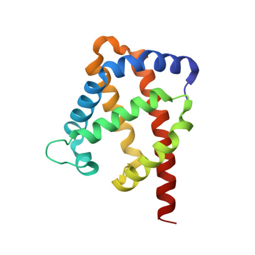
Crystallographic structure determination of B10 mutants of Vitreoscilla hemoglobin: role of Tyr29 (B10) in the structure of the ligand-binding site.

Ratakonda, S.Anand, A.Dikshit, K.Stark, B.C.Howard, A.J.

(2013) Acta Crystallogr Sect F Struct Biol Cryst Commun 69: 215-222

  • DOI: https://doi.org/10.1107/S1744309112044818
  • Primary Citation of Related Structures:  
    3TLD, 3TM3, 3TM9

  • PubMed Abstract: 

    Site-directed mutants of the gene encoding wild-type Vitreoscilla hemoglobin were made that changed Tyr29 (B10) of the wild-type Vitreoscilla hemoglobin (VHb) to either Phe or Ala. The wild-type and the two mutant hemoglobins were expressed in Escherichia coli and purified to homogeneity. The binding of the two mutants to CO was essentially identical to that of wild-type VHb as determined by CO-difference spectra. Circular-dichroism spectra also showed the two mutants to be essentially the same as wild-type VHb regarding overall helicity. All three VHbs were crystallized and their structures were determined at resolutions of 1.7-1.9 Å, which are similar to that of the original wild-type structure determination. The Tyr29Phe mutant has a structure that is essentially indistinguishable from that of the wild type. However, the structure of the Tyr29Ala mutant has significant differences from that of the wild type. In addition, for the Tyr29Ala mutant it was possible to determine the positions of most of the residues in the D region, which was disordered in the originally reported structure of wild-type VHb as well as in the wild-type VHb structure reported here. In the Tyr29Ala mutant, the five-membered ring of proline E8 (Pro54) occupies the space occupied by the aromatic ring of Tyr29 in the wild-type structure. These results are discussed in the context of the proposed role of Tyr29 in the structure of the oxygen-binding pocket.


  • Organizational Affiliation

    Biology Division, Department of Biological and Chemical Sciences, Illinois Institute of Technology, Chicago, IL 60616, USA.


Macromolecules
Find similar proteins by:  (by identity cutoff)  |  3D Structure
Entity ID: 1
MoleculeChains Sequence LengthOrganismDetailsImage
Bacterial hemoglobin146Vitreoscilla stercorariaMutation(s): 1 
Gene Names: vhb
UniProt
Find proteins for P04252 (Vitreoscilla stercoraria)
Explore P04252 
Go to UniProtKB:  P04252
Entity Groups  
Sequence Clusters30% Identity50% Identity70% Identity90% Identity95% Identity100% Identity
UniProt GroupP04252
Sequence Annotations
Expand
  • Reference Sequence
Small Molecules
Ligands 2 Unique
IDChains Name / Formula / InChI Key2D Diagram3D Interactions
HEM
Query on HEM

Download Ideal Coordinates CCD File 
B [auth A]PROTOPORPHYRIN IX CONTAINING FE
C34 H32 Fe N4 O4
KABFMIBPWCXCRK-RGGAHWMASA-L
EDO
Query on EDO

Download Ideal Coordinates CCD File 
C [auth A]
D [auth A]
E [auth A]
F [auth A]
G [auth A]
C [auth A],
D [auth A],
E [auth A],
F [auth A],
G [auth A],
H [auth A],
I [auth A],
J [auth A],
K [auth A],
L [auth A],
M [auth A],
N [auth A]
1,2-ETHANEDIOL
C2 H6 O2
LYCAIKOWRPUZTN-UHFFFAOYSA-N
Experimental Data & Validation

Experimental Data

  • Method: X-RAY DIFFRACTION
  • Resolution: 1.72 Å
  • R-Value Free: 0.206 
  • R-Value Work: 0.172 
  • R-Value Observed: 0.176 
  • Space Group: C 1 2 1
Unit Cell:
Length ( Å )Angle ( ˚ )
a = 85.194α = 90
b = 46.426β = 128.84
c = 62.873γ = 90
Software Package:
Software NamePurpose
PHENIXrefinement
PHASERphasing
REFMACrefinement
X-GENdata reduction
X-GENdata scaling
SERGUIdata collection

Structure Validation

View Full Validation Report


Ligand Structure Quality Assessment 


Entry History 

Deposition Data

Revision History  (Full details and data files)

  • Version 1.0: 2014-04-16
    Type: Initial release
  • Version 1.1: 2019-07-17
    Changes: Data collection, Refinement description
  • Version 1.2: 2024-02-28
    Changes: Data collection, Database references, Derived calculations