3TH9 | pdb_00003th9

Crystal Structure of HIV-1 Protease Mutant Q7K V32I L63I with a cyclic sulfonamide inhibitor


Experimental Data Snapshot

  • Method: X-RAY DIFFRACTION
  • Resolution: 1.34 Å
  • R-Value Free: 
    0.190 (Depositor), 0.187 (DCC) 
  • R-Value Work: 
    0.181 (Depositor) 
  • R-Value Observed: 
    0.181 (Depositor) 

wwPDB Validation 3D Report Full Report

Validation slider image for 3TH9

Ligand Structure Quality Assessment 


This is version 1.2 of the entry. See complete history

Literature

Design, Synthesis, and X-ray Crystallographic Analysis of a Novel Class of HIV-1 Protease Inhibitors.

Ganguly, A.K.Alluri, S.S.Caroccia, D.Biswas, D.Wang, C.H.Kang, E.Zhang, Y.McPhail, A.T.Carroll, S.S.Burlein, C.Munshi, V.Orth, P.Strickland, C.

(2011) J Med Chem 54: 7176-7183

  • DOI: https://doi.org/10.1021/jm200778q
  • Primary Citation Related Structures: 
    3TH9

  • PubMed Abstract: 

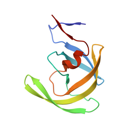
    In the present paper, design, synthesis, X-ray crystallographic analysis, and HIV-1 protease inhibitory activities of a novel class of compounds are disclosed. Compounds 28-30, 32, 35, and 40 were synthesized and found to be inhibitors of the HIV-1 protease. The crucial step in their synthesis involved an unusual endo radical cyclization process. Absolute stereochemistry of the three asymmetric centers in the above compounds have been established to be (4S,2'R,3'S) for optimal potency. X-ray crystallographic analysis has been used to determine the binding mode of the inhibitors to the HIV-1 protease.


  • Organizational Affiliation
    • Department of Chemistry, Chemical Biology and Biomedical Engineering, Stevens Institute of Technology, Hoboken, New Jersey 07030, United States. akganguly1@aol.com

Macromolecule Content 

  • Total Structure Weight: 22.59 kDa 
  • Atom Count: 1,813 
  • Modeled Residue Count: 198 
  • Deposited Residue Count: 198 
  • Unique protein chains: 1

Macromolecules

Find similar proteins by:|  3D Structure
Entity ID: 1
MoleculeChains  Sequence LengthOrganismDetailsImage
Gag-Pol polyprotein
A, B
99Human immunodeficiency virus type 1 lw12.3 isolateMutation(s): 3 
Gene Names: gag-pol
EC: 3.4.23.16
UniProt
Find proteins for P0C6F2 (Human immunodeficiency virus type 1 group M subtype B (isolate LW123))
Explore P0C6F2 
Go to UniProtKB:  P0C6F2
Entity Groups
Sequence Clusters30% Identity50% Identity70% Identity90% Identity95% Identity100% Identity
UniProt GroupP0C6F2
Sequence Annotations
Expand
Reference Sequence

Small Molecules

Ligands 1 Unique
IDChains Name / Formula / InChI Key2D Diagram3D Interactions
9Y9

Query on 9Y9


Download:Ideal Coordinates CCD File
C [auth A],
D [auth B]
tert-butyl {(2S,3R)-4-[(4S)-7-fluoro-4-methyl-1,1-dioxido-4,5-dihydro-1,2-benzothiazepin-2(3H)-yl]-3-hydroxy-1-phenylbutan-2-yl}carbamate
C25 H33 F N2 O5 S
KMJVCYPPUACVBA-BULFRSBZSA-N

Experimental Data & Validation

Experimental Data

  • Method: X-RAY DIFFRACTION
  • Resolution: 1.34 Å
  • R-Value Free:  0.190 (Depositor), 0.187 (DCC) 
  • R-Value Work:  0.181 (Depositor) 
  • R-Value Observed: 0.181 (Depositor) 
Space Group: P 21 21 2
Unit Cell:
Length ( Å )Angle ( ˚ )
a = 58.68α = 90
b = 85.89β = 90
c = 46.27γ = 90
Software Package:
Software NamePurpose
BUSTER-TNTrefinement
PDB_EXTRACTdata extraction
ADSCdata collection
XDSdata reduction
SCALAdata scaling
AMoREphasing
BUSTERrefinement

Structure Validation

View Full Validation Report


Ligand Structure Quality Assessment 


Entry History 

Deposition Data

  • Released Date: 2011-09-28 
  • Deposition Author(s): Orth, P.

Revision History  (Full details and data files)

  • Version 1.0: 2011-09-28
    Type: Initial release
  • Version 1.1: 2011-11-02
    Changes: Database references
  • Version 1.2: 2024-02-28
    Changes: Data collection, Database references, Derived calculations