3TFT | pdb_00003tft

Crystal structure of 7,8-diaminopelargonic acid synthase (BioA) from Mycobacterium tuberculosis, pre-reaction complex with a 3,6-dihydropyrid-2-one heterocycle inhibitor


Experimental Data Snapshot

  • Method: X-RAY DIFFRACTION
  • Resolution: 1.95 Å
  • R-Value Free: 
    0.197 (Depositor), 0.195 (DCC) 
  • R-Value Work: 
    0.156 (Depositor), 0.154 (DCC) 
  • R-Value Observed: 
    0.158 (Depositor) 

Starting Model: experimental
View more details

wwPDB Validation 3D Report Full Report

Validation slider image for 3TFT

Ligand Structure Quality Assessment 


This is version 1.2 of the entry. See complete history

Literature

Mechanism-based Inactivation by Aromatization of the Transaminase BioA Involved in Biotin Biosynthesis in Mycobaterium tuberculosis.

Shi, C.Geders, T.W.Park, S.W.Wilson, D.J.Boshoff, H.I.Abayomi, O.Barry, C.E.Schnappinger, D.Finzel, B.C.Aldrich, C.C.

(2011) J Am Chem Soc 133: 18194-18201

  • DOI: https://doi.org/10.1021/ja204036t
  • Primary Citation Related Structures: 
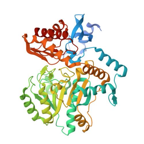
    3TFT, 3TFU

  • PubMed Abstract: 

    BioA catalyzes the second step of biotin biosynthesis, and this enzyme represents a potential target to develop new antitubercular agents. Herein we report the design, synthesis, and biochemical characterization of a mechanism-based inhibitor (1) featuring a 3,6-dihydropyrid-2-one heterocycle that covalently modifies the pyridoxal 5'-phosphate (PLP) cofactor of BioA through aromatization. The structure of the PLP adduct was confirmed by MS/MS and X-ray crystallography at 1.94 Å resolution. Inactivation of BioA by 1 was time- and concentration-dependent and protected by substrate. We used a conditional knock-down mutant of M. tuberculosis to demonstrate the antitubercular activity of 1 correlated with BioA expression, and these results provide support for the designed mechanism of action.


  • Organizational Affiliation
    • Academic Health Center, University of Minnesota, Minnesota 55455, United States.

Macromolecule Content 

  • Total Structure Weight: 97.65 kDa 
  • Atom Count: 7,239 
  • Modeled Residue Count: 846 
  • Deposited Residue Count: 914 
  • Unique protein chains: 1

Macromolecules

Find similar proteins by:|  3D Structure
Entity ID: 1
MoleculeChains  Sequence LengthOrganismDetailsImage
Adenosylmethionine-8-amino-7-oxononanoate aminotransferase
A, B
457Mycobacterium tuberculosisMutation(s): 0 
Gene Names: bioAMT1619MTCY336.35cRv1568
EC: 2.6.1.62
UniProt
Find proteins for P9WQ81 (Mycobacterium tuberculosis (strain ATCC 25618 / H37Rv))
Explore P9WQ81 
Go to UniProtKB:  P9WQ81
Entity Groups
Sequence Clusters30% Identity50% Identity70% Identity90% Identity95% Identity100% Identity
UniProt GroupP9WQ81
Sequence Annotations
Expand
Reference Sequence

Experimental Data & Validation

Experimental Data

  • Method: X-RAY DIFFRACTION
  • Resolution: 1.95 Å
  • R-Value Free:  0.197 (Depositor), 0.195 (DCC) 
  • R-Value Work:  0.156 (Depositor), 0.154 (DCC) 
  • R-Value Observed: 0.158 (Depositor) 
Space Group: P 21 21 21
Unit Cell:
Length ( Å )Angle ( ˚ )
a = 63.057α = 90
b = 66.385β = 90
c = 203.13γ = 90
Software Package:
Software NamePurpose
d*TREKdata scaling
d*TREKdata reduction
PHASERphasing
PHENIXrefinement
PDB_EXTRACTdata extraction
Blu-Icedata collection

Structure Validation

View Full Validation Report


Ligand Structure Quality Assessment 


Entry History 

Deposition Data

Revision History  (Full details and data files)

  • Version 1.0: 2011-10-19
    Type: Initial release
  • Version 1.1: 2011-11-23
    Changes: Database references
  • Version 1.2: 2023-09-13
    Changes: Data collection, Database references, Derived calculations, Refinement description