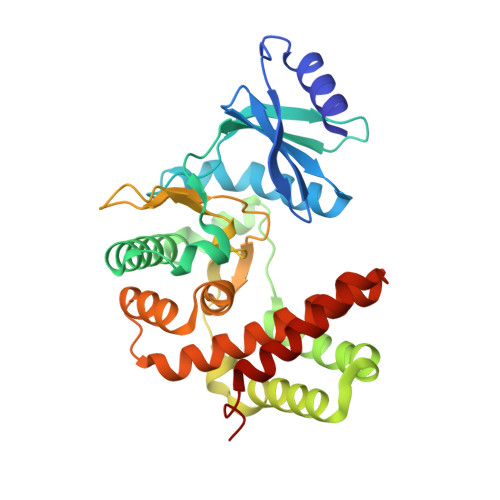
Aminoglycoside 2''-Phosphotransferase IIIa (APH(2'')-IIIa) Prefers GTP over ATP: STRUCTURAL TEMPLATES FOR NUCLEOTIDE RECOGNITION IN THE BACTERIAL AMINOGLYCOSIDE-2'' KINASES.
Smith, C.A., Toth, M., Frase, H., Byrnes, L.J., Vakulenko, S.B.(2012) J Biological Chem 287: 12893-12903
- PubMed: 22367198
- DOI: https://doi.org/10.1074/jbc.M112.341206
- Primary Citation of Related Structures:
3TDV, 3TDW - PubMed Abstract:
Contrary to the accepted dogma that ATP is the canonical phosphate donor in aminoglycoside kinases and protein kinases, it was recently demonstrated that all members of the bacterial aminoglycoside 2''-phosphotransferase IIIa (APH(2'')) aminoglycoside kinase family are unique in their ability to utilize GTP as a cofactor for antibiotic modification. Here we describe the structural determinants for GTP recognition in these enzymes. The crystal structure of the GTP-dependent APH(2'')-IIIa shows that although this enzyme has templates for both ATP and GTP binding superimposed on a single nucleotide specificity motif, access to the ATP-binding template is blocked by a bulky tyrosine residue. Substitution of this tyrosine by a smaller amino acid opens access to the ATP template. Similar GTP binding templates are conserved in other bacterial aminoglycoside kinases, whereas in the structurally related eukaryotic protein kinases this template is less conserved. The aminoglycoside kinases are important antibiotic resistance enzymes in bacteria, whose wide dissemination severely limits available therapeutic options, and the GTP binding templates could be exploited as new, previously unexplored targets for inhibitors of these clinically important enzymes.
- Stanford Synchrotron Radiation Lightsource, Stanford University, Menlo Park, California 94025, USA. csmith@slac.stanford.edu
Organizational Affiliation:


