


Crystal structure of the complex of Dihydrodipicolinate synthase from Acinetobacter baumannii with 2-Ketobutanoic acid at 1.99 A resolution
Kumar, M., Kaushik, S., Sinha, M., Kaur, P., Tewari, R., Sharma, S., Singh, T.P.To be published.
Experimental Data Snapshot
Starting Model: experimental
View more details
wwPDB Validation 3D Report Full Report

Entity ID: 1 | |||||
|---|---|---|---|---|---|
| Molecule | Chains | Sequence Length | Organism | Details | Image |
| Dihydrodipicolinate synthase | 291 | Acinetobacter baumannii ATCC 19606 = CIP 70.34 = JCM 6841 | Mutation(s): 0 Gene Names: dhdps, HMPREF0010_03414 EC: 4.2.1.52 (PDB Primary Data), 4.3.3.7 (UniProt) | 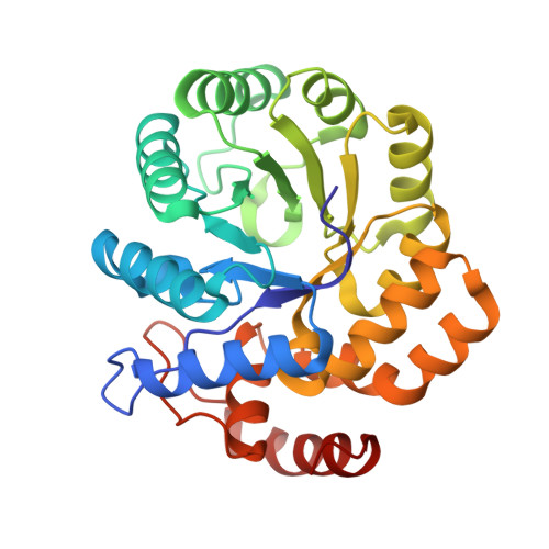 | |
UniProt | |||||
Entity Groups | |||||
| Sequence Clusters | 30% Identity50% Identity70% Identity90% Identity95% Identity100% Identity | ||||
| UniProt Group | D0CFC3 | ||||
Sequence AnnotationsExpand | |||||
Reference Sequence | |||||
| Ligands 1 Unique | |||||
|---|---|---|---|---|---|
| ID | Chains | Name / Formula / InChI Key | 2D Diagram | 3D Interactions | |
| 2KT Download:Ideal Coordinates CCD File | C [auth A], D [auth B] | 2-KETOBUTYRIC ACID C4 H6 O3 TYEYBOSBBBHJIV-UHFFFAOYSA-N |  | ||
| Length ( Å ) | Angle ( ˚ ) |
|---|---|
| a = 52.235 | α = 90 |
| b = 121.947 | β = 115.9 |
| c = 51.131 | γ = 90 |
| Software Name | Purpose |
|---|---|
| HKL-2000 | data collection |
| AMoRE | phasing |
| REFMAC | refinement |
| HKL-2000 | data reduction |
| SCALEPACK | data scaling |


