3TBV | pdb_00003tbv

CRYSTAL STRUCTURE OF THE MURINE CLASS I MAJOR HISTOCOMPATIBILITY COMPLEX H-2DB IN COMPLEX WITH THE LCMV-DERIVED GP33 ALTERED PEPTIDE ligand (A2G,V3P,Y4A)


Experimental Data Snapshot

  • Method: X-RAY DIFFRACTION
  • Resolution: 2.10 Å
  • R-Value Free: 
    0.278 (Depositor), 0.282 (DCC) 
  • R-Value Work: 
    0.225 (Depositor), 0.233 (DCC) 
  • R-Value Observed: 
    0.227 (Depositor) 

Starting Model: experimental
View more details

wwPDB Validation 3D Report Full Report

Validation slider image for 3TBV

This is version 1.2 of the entry. See complete history

Literature

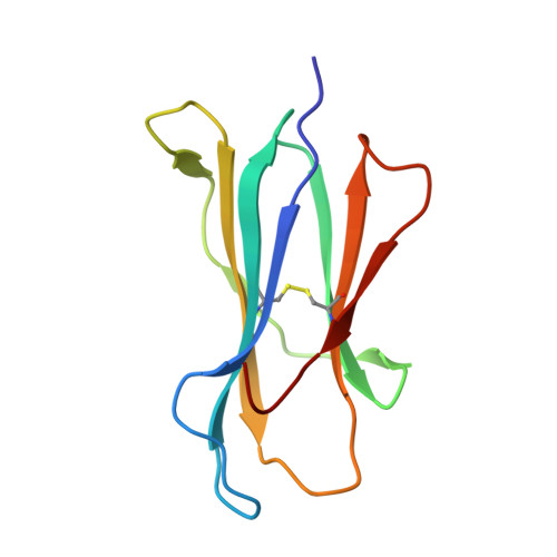
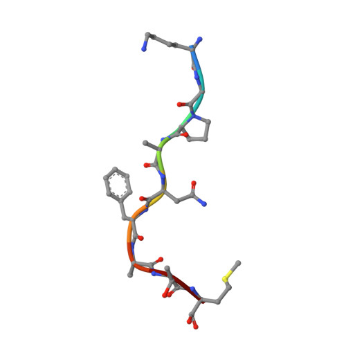
Conversion of a T cell viral antagonist into an agonist through higher stabilization and conserved molecular mimicry: Implications for TCR recognition

Duru, A.D.Allerbring, E.B.Uchtenhagen, H.Mazumdar, P.A.Badia-Martinez, D.Madhurantakam, C.Sandalova, T.Nygren, P.Achour, A.

To be published.

Macromolecule Content 

  • Total Structure Weight: 179.95 kDa 
  • Atom Count: 13,548 
  • Modeled Residue Count: 1,536 
  • Deposited Residue Count: 1,536 
  • Unique protein chains: 3

Macromolecules

Find similar proteins by:|  3D Structure
Entity ID: 1
MoleculeChains  Sequence LengthOrganismDetailsImage
H-2 class I histocompatibility antigen, D-B alpha chain
A, C, E, G
276Mus musculusMutation(s): 0 
Gene Names: H2-D1H2-DB
UniProt
Find proteins for P01899 (Mus musculus)
Explore P01899 
Go to UniProtKB:  P01899
Entity Groups
Sequence Clusters30% Identity50% Identity70% Identity90% Identity95% Identity100% Identity
UniProt GroupP01899
Sequence Annotations
Expand
Reference Sequence
Find similar proteins by:|  3D Structure
Entity ID: 2
MoleculeChains  Sequence LengthOrganismDetailsImage
Beta-2-microglobulin
B, D, F, H
99Mus musculusMutation(s): 0 
Gene Names: B2m
UniProt & NIH Common Fund Data Resources
Find proteins for P01887 (Mus musculus)
Explore P01887 
Go to UniProtKB:  P01887
IMPC:  MGI:88127
Entity Groups
Sequence Clusters30% Identity50% Identity70% Identity90% Identity95% Identity100% Identity
UniProt GroupP01887
Sequence Annotations
Expand
Reference Sequence
Find similar proteins by:  Sequence   |   3D Structure  
Entity ID: 3
MoleculeChains  Sequence LengthOrganismDetailsImage
Glycoprotein G1
I, J, K, L
9Lymphocytic choriomeningitis virus (strain WE)Mutation(s): 4 
UniProt
Find proteins for P07399 (Lymphocytic choriomeningitis virus (strain WE))
Explore P07399 
Go to UniProtKB:  P07399
Entity Groups
UniProt GroupP07399
Sequence Annotations
Expand
Reference Sequence

Small Molecules

Ligands 2 Unique
IDChains Name / Formula / InChI Key2D Diagram3D Interactions
SO4

Query on SO4


Download:Ideal Coordinates CCD File
M [auth A],
Q [auth C],
R [auth E],
U [auth F]
SULFATE ION
O4 S
QAOWNCQODCNURD-UHFFFAOYSA-L
GOL

Query on GOL


Download:Ideal Coordinates CCD File
N [auth A]
O [auth A]
P [auth B]
S [auth E]
T [auth E]
N [auth A],
O [auth A],
P [auth B],
S [auth E],
T [auth E],
V [auth H],
W [auth H]
GLYCEROL
C3 H8 O3
PEDCQBHIVMGVHV-UHFFFAOYSA-N

Experimental Data & Validation

Experimental Data

  • Method: X-RAY DIFFRACTION
  • Resolution: 2.10 Å
  • R-Value Free:  0.278 (Depositor), 0.282 (DCC) 
  • R-Value Work:  0.225 (Depositor), 0.233 (DCC) 
  • R-Value Observed: 0.227 (Depositor) 
Space Group: P 1 21 1
Unit Cell:
Length ( Å )Angle ( ˚ )
a = 96.584α = 90
b = 126.473β = 106.71
c = 102.11γ = 90
Software Package:
Software NamePurpose
ADSCdata collection
PHASERphasing
PHENIXrefinement
MOSFLMdata reduction
SCALAdata scaling

Structure Validation

View Full Validation Report


Entry History 

Deposition Data

Revision History  (Full details and data files)

  • Version 1.0: 2012-08-08
    Type: Initial release
  • Version 1.1: 2023-09-13
    Changes: Data collection, Database references, Derived calculations, Refinement description
  • Version 1.2: 2024-11-27
    Changes: Structure summary