3T0W

Fluorogen activating protein M8VL in complex with dimethylindole red


Experimental Data Snapshot

  • Method: X-RAY DIFFRACTION
  • Resolution: 1.50 Å
  • R-Value Free: 0.183 
  • R-Value Work: 0.148 
  • R-Value Observed: 0.150 

wwPDB Validation   3D Report Full Report


Ligand Structure Quality Assessment 


This is version 2.0 of the entry. See complete history


Literature

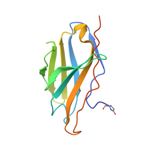
A variable light domain fluorogen activating protein homodimerizes to activate dimethylindole red.

Senutovitch, N.Stanfield, R.L.Bhattacharyya, S.Rule, G.S.Wilson, I.A.Armitage, B.A.Waggoner, A.S.Berget, P.B.

(2012) Biochemistry 51: 2471-2485

  • DOI: https://doi.org/10.1021/bi201422g
  • Primary Citation of Related Structures:  
    3T0V, 3T0W, 3T0X

  • PubMed Abstract: 

    Novel fluorescent tools such as green fluorescent protein analogues and fluorogen activating proteins (FAPs) are useful in biological imaging for tracking protein dynamics in real time with a low fluorescence background. FAPs are single-chain variable fragments (scFvs) selected from a yeast surface display library that produce fluorescence upon binding a specific dye or fluorogen that is normally not fluorescent when present in solution. FAPs generally consist of human immunoglobulin variable heavy (V(H)) and variable light (V(L)) domains covalently attached via a glycine- and serine-rich linker. Previously, we determined that the yeast surface clone, V(H)-V(L) M8, could bind and activate the fluorogen dimethylindole red (DIR) but that the fluorogen activation properties were localized to the M8V(L) domain. We report here that both nuclear magnetic resonance and X-ray diffraction methods indicate the M8V(L) forms noncovalent, antiparallel homodimers that are the fluorogen activating species. The M8V(L) homodimers activate DIR by restriction of internal rotation of the bound dye. These structural results, together with directed evolution experiments with both V(H)-V(L) M8 and M8V(L), led us to rationally design tandem, covalent homodimers of M8V(L) domains joined by a flexible linker that have a high affinity for DIR and good quantum yields.


  • Organizational Affiliation

    The Department of Biological Sciences, Carnegie Mellon University, Pittsburgh, Pennsylvania 15213, United States.


Macromolecules
Find similar proteins by:  (by identity cutoff)  |  3D Structure
Entity ID: 1
MoleculeChains Sequence LengthOrganismDetailsImage
immunoglobulin variable lambda domain
A, B
123Homo sapiensMutation(s): 0 
Entity Groups  
Sequence Clusters30% Identity50% Identity70% Identity90% Identity95% Identity100% Identity
Sequence Annotations
Expand
  • Reference Sequence
Small Molecules
Ligands 3 Unique
IDChains Name / Formula / InChI Key2D Diagram3D Interactions
PE3
Query on PE3

Download Ideal Coordinates CCD File 
E [auth A]3,6,9,12,15,18,21,24,27,30,33,36,39-TRIDECAOXAHENTETRACONTANE-1,41-DIOL
C28 H58 O15
ILLKMACMBHTSHP-UHFFFAOYSA-N
DIW
Query on DIW

Download Ideal Coordinates CCD File 
F [auth A]1-(3-sulfopropyl)-4-[(1E,3E)-3-(1,3,3-trimethyl-1,3-dihydro-2H-indol-2-ylidene)prop-1-en-1-yl]quinolinium
C26 H29 N2 O3 S
NTABMUJQZABQGD-UHFFFAOYSA-O
CL
Query on CL

Download Ideal Coordinates CCD File 
C [auth A],
D [auth A]
CHLORIDE ION
Cl
VEXZGXHMUGYJMC-UHFFFAOYSA-M
Modified Residues  1 Unique
IDChains TypeFormula2D DiagramParent
PCA
Query on PCA
A, B
L-PEPTIDE LINKINGC5 H7 N O3GLN
Binding Affinity Annotations 
IDSourceBinding Affinity
DIW PDBBind:  3T0W Kd: 50 (nM) from 1 assay(s)
Experimental Data & Validation

Experimental Data

  • Method: X-RAY DIFFRACTION
  • Resolution: 1.50 Å
  • R-Value Free: 0.183 
  • R-Value Work: 0.148 
  • R-Value Observed: 0.150 
  • Space Group: P 21 21 2
Unit Cell:
Length ( Å )Angle ( ˚ )
a = 73.899α = 90
b = 83.533β = 90
c = 35.142γ = 90
Software Package:
Software NamePurpose
Blu-Icedata collection
PHASERphasing
PHENIXrefinement
HKL-2000data reduction
SCALEPACKdata scaling

Structure Validation

View Full Validation Report


Ligand Structure Quality Assessment 


Entry History 

Deposition Data

Revision History  (Full details and data files)

  • Version 1.0: 2012-03-21
    Type: Initial release
  • Version 1.1: 2012-03-28
    Changes: Other
  • Version 1.2: 2012-04-11
    Changes: Database references
  • Version 2.0: 2019-12-25
    Changes: Derived calculations, Polymer sequence