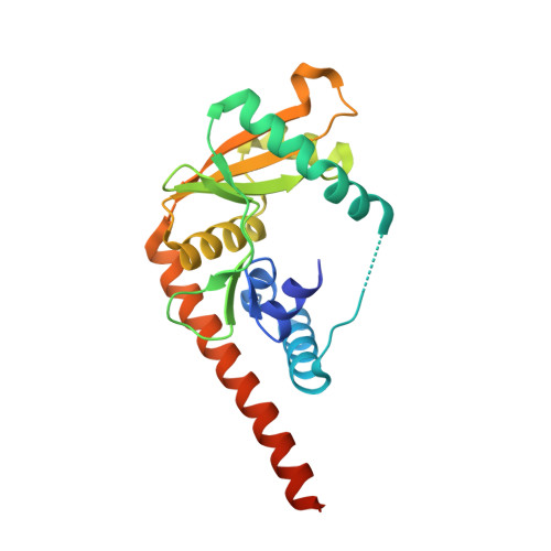
Structural and functional insight into the mechanism of an alkaline exonuclease from Laribacter hongkongensis.
Yang, W., Chen, W.Y., Wang, H., Ho, J.W., Huang, J.D., Woo, P.C., Lau, S.K., Yuen, K.Y., Zhang, Q., Zhou, W., Bartlam, M., Watt, R.M., Rao, Z.(2011) Nucleic Acids Res 39: 9803-9819
- PubMed: 21893587 Search on PubMedSearch on PubMed Central
- DOI: https://doi.org/10.1093/nar/gkr660
- Primary Citation Related Structures:
3SYY, 3SZ4, 3SZ5 - PubMed Abstract:
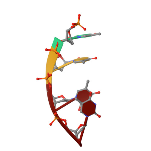
Alkaline exonuclease and single-strand DNA (ssDNA) annealing proteins (SSAPs) are key components of DNA recombination and repair systems within many prokaryotes, bacteriophages and virus-like genetic elements. The recently sequenced β-proteobacterium Laribacter hongkongensis (strain HLHK9) encodes putative homologs of alkaline exonuclease (LHK-Exo) and SSAP (LHK-Bet) proteins on its 3.17 Mb genome. Here, we report the biophysical, biochemical and structural characterization of recombinant LHK-Exo protein. LHK-Exo digests linear double-stranded DNA molecules from their 5'-termini in a highly processive manner. Exonuclease activities are optimum at pH 8.2 and essentially require Mg(2+) or Mn(2+) ions. 5'-phosphorylated DNA substrates are preferred over dephosphorylated ones. The crystal structure of LHK-Exo was resolved to 1.9 Å, revealing a 'doughnut-shaped' toroidal trimeric arrangement with a central tapered channel, analogous to that of λ-exonuclease (Exo) from bacteriophage-λ. Active sites containing two bound Mg(2+) ions on each of the three monomers were located in clefts exposed to this central channel. Crystal structures of LHK-Exo in complex with dAMP and ssDNA were determined to elucidate the structural basis for substrate recognition and binding. Through structure-guided mutational analysis, we discuss the roles played by various active site residues. A conserved two metal ion catalytic mechanism is proposed for this class of alkaline exonucleases.
- State Key Laboratory of Medicinal Chemical Biology, Nankai University, Tianjin 300071, China.
Organizational Affiliation: