3ST5 | pdb_00003st5

Crystal structure of wild-type HIV-1 protease with C3-Substituted Hexahydrocyclopentafuranyl Urethane as P2-Ligand, GRL-0489A


Experimental Data Snapshot

  • Method: X-RAY DIFFRACTION
  • Resolution: 1.45 Å
  • R-Value Free: 
    0.219 (Depositor), 0.211 (DCC) 
  • R-Value Work: 
    0.157 (Depositor), 0.163 (DCC) 
  • R-Value Observed: 
    0.157 (Depositor) 

Starting Model: experimental
View more details

wwPDB Validation 3D Report Full Report

Validation slider image for 3ST5

Ligand Structure Quality Assessment 


This is version 1.3 of the entry. See complete history

Literature

Design of HIV-1 Protease Inhibitors with C3-Substituted Hexahydrocyclopentafuranyl Urethanes as P2-Ligands: Synthesis, Biological Evaluation, and Protein-Ligand X-ray Crystal Structure.

Ghosh, A.K.Chapsal, B.D.Parham, G.L.Steffey, M.Agniswamy, J.Wang, Y.F.Amano, M.Weber, I.T.Mitsuya, H.

(2011) J Med Chem 54: 5890-5901

  • DOI: https://doi.org/10.1021/jm200649p
  • Primary Citation Related Structures: 
    3ST5

  • PubMed Abstract: 

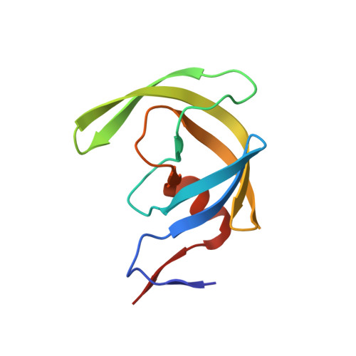
    We report the design, synthesis, biological evaluation, and the X-ray crystal structure of a novel inhibitor bound to the HIV-1 protease. Various C3-functionalized cyclopentanyltetrahydrofurans (Cp-THF) were designed to interact with the flap Gly48 carbonyl or amide NH in the S2-subsite of the HIV-1 protease. We investigated the potential of those functionalized ligands in combination with hydroxyethylsulfonamide isosteres. Inhibitor 26 containing a 3-(R)-hydroxyl group on the Cp-THF core displayed the most potent enzyme inhibitory and antiviral activity. Our studies revealed a preference for the 3-(R)-configuration over the corresponding 3-(S)-derivative. Inhibitor 26 exhibited potent activity against a panel of multidrug-resistant HIV-1 variants. A high resolution X-ray structure of 26-bound HIV-1 protease revealed important molecular insight into the ligand-binding site interactions.


  • Organizational Affiliation
    • Department of Chemistry, Purdue University, West Lafayette, Indiana 47907, USA. akghosh@purdue.edu

Macromolecule Content 

  • Total Structure Weight: 22.13 kDa 
  • Atom Count: 1,874 
  • Modeled Residue Count: 198 
  • Deposited Residue Count: 198 
  • Unique protein chains: 1

Macromolecules

Find similar proteins by:|  3D Structure
Entity ID: 1
MoleculeChains  Sequence LengthOrganismDetailsImage
Protease
A, B
99Human immunodeficiency virus type 1 (BRU ISOLATE)Mutation(s): 5 
Gene Names: gag-pol
EC: 3.4.23.16
UniProt
Find proteins for P03367 (Human immunodeficiency virus type 1 group M subtype B (isolate BRU/LAI))
Explore P03367 
Go to UniProtKB:  P03367
Entity Groups
Sequence Clusters30% Identity50% Identity70% Identity90% Identity95% Identity100% Identity
UniProt GroupP03367
Sequence Annotations
Expand
Reference Sequence

Small Molecules

Ligands 2 Unique
IDChains Name / Formula / InChI Key2D Diagram3D Interactions
G89

Query on G89


Download:Ideal Coordinates CCD File
D [auth B](3R,3aR,5R,6aR)-3-hydroxyhexahydro-2H-cyclopenta[b]furan-5-yl [(2S,3R)-3-hydroxy-4-{[(4-methoxyphenyl)sulfonyl](2-methylpropyl)amino}-1-phenylbutan-2-yl]carbamate
C29 H40 N2 O8 S
LJPOEDMTSRUIGG-RVSUYJHDSA-N
CL

Query on CL


Download:Ideal Coordinates CCD File
C [auth A],
E [auth B]
CHLORIDE ION
Cl
VEXZGXHMUGYJMC-UHFFFAOYSA-M

Experimental Data & Validation

Experimental Data

  • Method: X-RAY DIFFRACTION
  • Resolution: 1.45 Å
  • R-Value Free:  0.219 (Depositor), 0.211 (DCC) 
  • R-Value Work:  0.157 (Depositor), 0.163 (DCC) 
  • R-Value Observed: 0.157 (Depositor) 
Space Group: P 21 21 2
Unit Cell:
Length ( Å )Angle ( ˚ )
a = 58.39α = 90
b = 86.552β = 90
c = 45.846γ = 90
Software Package:
Software NamePurpose
HKL-2000data collection
PHASESphasing
SHELXL-97refinement
HKL-2000data reduction
HKL-2000data scaling

Structure Validation

View Full Validation Report


Ligand Structure Quality Assessment 


Entry History 

Deposition Data

Revision History  (Full details and data files)

  • Version 1.0: 2011-08-17
    Type: Initial release
  • Version 1.1: 2011-08-31
    Changes: Database references
  • Version 1.2: 2012-12-12
    Changes: Other
  • Version 1.3: 2023-09-13
    Changes: Data collection, Database references, Derived calculations, Refinement description