


Crystal structure of L-Azetidine-2-carboxylate hydrolase from Pseudomonas sp. strain A2C
Jitsumori, K., Toyoda, M., Mikami, B., Wackett, L.P., Kurihara, T., Esaki, N.To be published.
Experimental Data Snapshot
wwPDB Validation 3D Report Full Report

Entity ID: 1 | |||||
|---|---|---|---|---|---|
| Molecule | Chains | Sequence Length | Organism | Details | Image |
| (S)-2-haloacid dehalogenase | 240 | Pseudomonas sp. A2C | Mutation(s): 0 EC: 3.8.1.2 (PDB Primary Data), 3.5 (UniProt) | 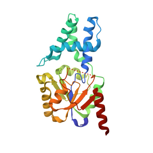 | |
UniProt | |||||
Entity Groups | |||||
| Sequence Clusters | 30% Identity50% Identity70% Identity90% Identity95% Identity100% Identity | ||||
| UniProt Group | B2Z3V8 | ||||
Sequence AnnotationsExpand | |||||
Reference Sequence | |||||
| Ligands 2 Unique | |||||
|---|---|---|---|---|---|
| ID | Chains | Name / Formula / InChI Key | 2D Diagram | 3D Interactions | |
| GOL Download:Ideal Coordinates CCD File | B [auth A] C [auth A] D [auth A] E [auth A] F [auth A] | GLYCEROL C3 H8 O3 PEDCQBHIVMGVHV-UHFFFAOYSA-N |  | ||
| IMD Download:Ideal Coordinates CCD File | K [auth A] | IMIDAZOLE C3 H5 N2 RAXXELZNTBOGNW-UHFFFAOYSA-O |  | ||
| Length ( Å ) | Angle ( ˚ ) |
|---|---|
| a = 35.612 | α = 90 |
| b = 63.588 | β = 105.54 |
| c = 54.665 | γ = 90 |
| Software Name | Purpose |
|---|---|
| HKL-2000 | data collection |
| PHENIX | model building |
| PHENIX | refinement |
| DENZO | data reduction |
| SCALEPACK | data scaling |
| PHENIX | phasing |


