3SMA

A new N-acetyltransferase fold in the structure and mechanism of the phosphonate biosynthetic enzyme FrbF


Experimental Data Snapshot

  • Method: X-RAY DIFFRACTION
  • Resolution: 2.00 Å
  • R-Value Free: 0.246 
  • R-Value Work: 0.193 
  • R-Value Observed: 0.195 

wwPDB Validation   3D Report Full Report


Ligand Structure Quality Assessment 


This is version 1.3 of the entry. See complete history


Literature

New N-Acetyltransferase Fold in the Structure and Mechanism of the Phosphonate Biosynthetic Enzyme FrbF.

Bae, B.Cobb, R.E.Desieno, M.A.Zhao, H.Nair, S.K.

(2011) J Biol Chem 286: 36132-36141

  • DOI: https://doi.org/10.1074/jbc.M111.263533
  • Primary Citation of Related Structures:  
    3SMA

  • PubMed Abstract: 

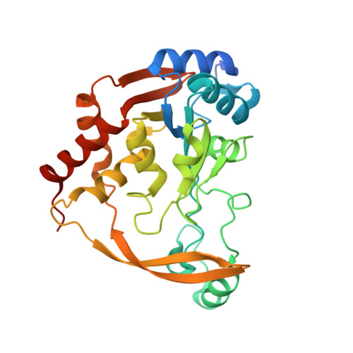
    The enzyme FrbF from Streptomyces rubellomurinus has attracted significant attention due to its role in the biosynthesis of the antimalarial phosphonate FR-900098. The enzyme catalyzes acetyl transfer onto the hydroxamate of the FR-900098 precursors cytidine 5'-monophosphate-3-aminopropylphosphonate and cytidine 5'-monophosphate-N-hydroxy-3-aminopropylphosphonate. Despite the established function as a bona fide N-acetyltransferase, FrbF shows no sequence similarity to any member of the GCN5-like N-acetyltransferase (GNAT) superfamily. Here, we present the 2.0 Å resolution crystal structure of FrbF in complex with acetyl-CoA, which demonstrates a unique architecture that is distinct from those of canonical GNAT-like acetyltransferases. We also utilized the co-crystal structure to guide structure-function studies that identified the roles of putative active site residues in the acetyltransferase mechanism. The combined biochemical and structural analyses of FrbF provide insights into this previously uncharacterized family of N-acetyltransferases and also provide a molecular framework toward the production of novel N-acyl derivatives of FR-900098.


  • Organizational Affiliation

    Department of Biochemistry, University of Illinois at Urbana-Champaign, Urbana, Illinois 61801.


Macromolecules
Find similar proteins by:  (by identity cutoff)  |  3D Structure
Entity ID: 1
MoleculeChains Sequence LengthOrganismDetailsImage
FrbF
A, B, C, D
286Streptomyces rubellomurinusMutation(s): 0 
Gene Names: frbF
UniProt
Find proteins for Q0ZQ43 (Streptomyces rubellomurinus (strain ATCC 31215))
Explore Q0ZQ43 
Go to UniProtKB:  Q0ZQ43
Entity Groups  
Sequence Clusters30% Identity50% Identity70% Identity90% Identity95% Identity100% Identity
UniProt GroupQ0ZQ43
Sequence Annotations
Expand
  • Reference Sequence
Experimental Data & Validation

Experimental Data

  • Method: X-RAY DIFFRACTION
  • Resolution: 2.00 Å
  • R-Value Free: 0.246 
  • R-Value Work: 0.193 
  • R-Value Observed: 0.195 
  • Space Group: P 1 2 1
Unit Cell:
Length ( Å )Angle ( ˚ )
a = 128.832α = 90
b = 35.692β = 117.6
c = 136.087γ = 90
Software Package:
Software NamePurpose
HKL-2000data collection
BUSTERrefinement
MOSFLMdata reduction
SCALAdata scaling

Structure Validation

View Full Validation Report


Ligand Structure Quality Assessment 


Entry History 

Deposition Data

Revision History  (Full details and data files)

  • Version 1.0: 2011-08-24
    Type: Initial release
  • Version 1.1: 2011-09-07
    Changes: Database references
  • Version 1.2: 2011-10-26
    Changes: Database references
  • Version 1.3: 2024-02-28
    Changes: Data collection, Database references, Derived calculations