3SGB | pdb_00003sgb

STRUCTURE OF THE COMPLEX OF STREPTOMYCES GRISEUS PROTEASE B AND THE THIRD DOMAIN OF THE TURKEY OVOMUCOID INHIBITOR AT 1.8 ANGSTROMS RESOLUTION


Experimental Data Snapshot

  • Method: X-RAY DIFFRACTION
  • Resolution: 1.80 Å
  • R-Value Work: 
    0.125 (Depositor) 

wwPDB Validation 3D Report Full Report

Validation slider image for 3SGB

This is version 1.4 of the entry. See complete history

Literature

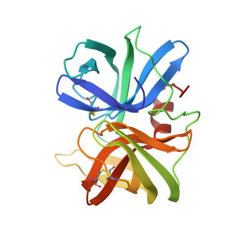
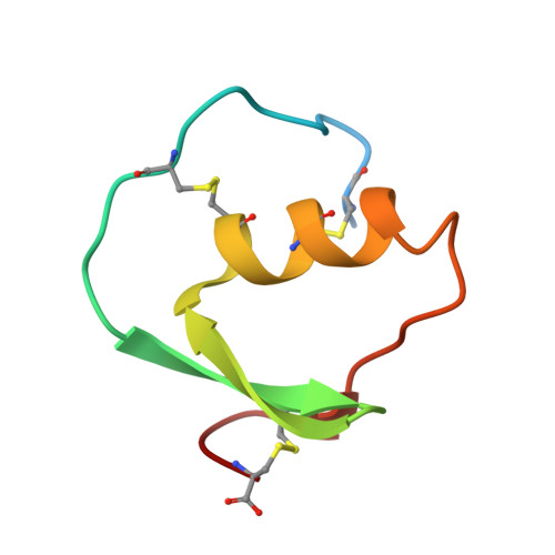
Structure of the complex of Streptomyces griseus protease B and the third domain of the turkey ovomucoid inhibitor at 1.8-A resolution.

Read, R.J.Fujinaga, M.Sielecki, A.R.James, M.N.

(1983) Biochemistry 22: 4420-4433

  • DOI: https://doi.org/10.1021/bi00288a012
  • Primary Citation Related Structures: 
    3SGB

  • PubMed Abstract: 

    The structure of the complex between the serine protease Streptomyces griseus protease B (SGPB) and the third domain of the Kazal-type ovomucoid inhibitor from turkey has been solved at 1.8-A resolution and refined to a conventional R factor of 0.125. As others have reported previously for analogous complexes of proteases and protein inhibitors, the inhibitor binds in a fashion similar to that of a substrate; it is not cleaved, but there is a close approach (2.7 A) of the active site nucleophile Ser-195 O gamma to the carbonyl carbon of the reactive peptide bond of the inhibitor. Contrary to the structural reports regarding the other enzyme-inhibitor complexes, we conclude that there is no evidence for a significant distortion of this peptide bond from planarity. The mechanism of inhibition can be understood in terms of the equilibrium thermodynamic parameters Ka, the enzyme-inhibitor association constant, and Khyd, the equilibrium constant for inhibitor hydrolysis. These thermodynamic parameters can be rationalized in terms of the observed structure.

Macromolecule Content 

  • Total Structure Weight: 24.69 kDa 
  • Atom Count: 1,872 
  • Modeled Residue Count: 235 
  • Deposited Residue Count: 241 
  • Unique protein chains: 2

Macromolecules

Find similar proteins by:|  3D Structure
Entity ID: 1
MoleculeChains  Sequence LengthOrganismDetailsImage
PROTEINASE B (SGPB)A [auth E]185Streptomyces griseusMutation(s): 0 
EC: 3.4.21.81
UniProt
Find proteins for P00777 (Streptomyces griseus)
Explore P00777 
Go to UniProtKB:  P00777
Entity Groups
Sequence Clusters30% Identity50% Identity70% Identity90% Identity95% Identity100% Identity
UniProt GroupP00777
Sequence Annotations
Expand
Reference Sequence
Find similar proteins by:|  3D Structure
Entity ID: 2
MoleculeChains  Sequence LengthOrganismDetailsImage
TURKEY OVOMUCOID INHIBITOR (OMTKY3)B [auth I]56Meleagris gallopavoMutation(s): 0 
UniProt
Find proteins for P68390 (Meleagris gallopavo)
Explore P68390 
Go to UniProtKB:  P68390
Entity Groups
Sequence Clusters30% Identity50% Identity70% Identity90% Identity95% Identity100% Identity
UniProt GroupP68390
Sequence Annotations
Expand
Reference Sequence

Experimental Data & Validation

Experimental Data

  • Method: X-RAY DIFFRACTION
  • Resolution: 1.80 Å
  • R-Value Work:  0.125 (Depositor) 
Space Group: P 1 21 1
Unit Cell:
Length ( Å )Angle ( ˚ )
a = 45.35α = 90
b = 54.52β = 119.2
c = 45.65γ = 90

Structure Validation

View Full Validation Report


Entry History 

Deposition Data

Revision History  (Full details and data files)

  • Version 1.0: 1983-07-12
    Type: Initial release
  • Version 1.1: 2008-03-25
    Changes: Version format compliance
  • Version 1.2: 2011-07-13
    Changes: Version format compliance
  • Version 1.3: 2017-11-29
    Changes: Derived calculations, Other
  • Version 1.4: 2024-10-30
    Changes: Data collection, Database references, Structure summary