3SBO

Structure of E.coli GDH from native source


Experimental Data Snapshot

  • Method: X-RAY DIFFRACTION
  • Resolution: 3.20 Å
  • R-Value Free: 0.265 
  • R-Value Work: 0.229 
  • R-Value Observed: 0.231 

wwPDB Validation   3D Report Full Report


This is version 1.3 of the entry. See complete history


Literature

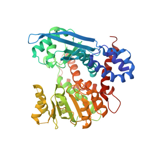
Macro-to-Micro Structural Proteomics: Native Source Proteins for High-Throughput Crystallization.

Totir, M.Echols, N.Nanao, M.Gee, C.L.Moskaleva, A.Gradia, S.Iavarone, A.T.Berger, J.M.May, A.P.Zubieta, C.Alber, T.

(2012) PLoS One 7: e32498-e32498

  • DOI: https://doi.org/10.1371/journal.pone.0032498
  • Primary Citation of Related Structures:  
    2XHY, 3N6Q, 3NBU, 3SBO

  • PubMed Abstract: 

    Structural biology and structural genomics projects routinely rely on recombinantly expressed proteins, but many proteins and complexes are difficult to obtain by this approach. We investigated native source proteins for high-throughput protein crystallography applications. The Escherichia coli proteome was fractionated, purified, crystallized, and structurally characterized. Macro-scale fermentation and fractionation were used to subdivide the soluble proteome into 408 unique fractions of which 295 fractions yielded crystals in microfluidic crystallization chips. Of the 295 crystals, 152 were selected for optimization, diffraction screening, and data collection. Twenty-three structures were determined, four of which were novel. This study demonstrates the utility of native source proteins for high-throughput crystallography.


  • Organizational Affiliation

    Department of Molecular and Cell Biology, University of California, Berkeley, California, United States of America.


Macromolecules
Find similar proteins by:  (by identity cutoff)  |  3D Structure
Entity ID: 1
MoleculeChains Sequence LengthOrganismDetailsImage
NADP-specific glutamate dehydrogenase
A, B, C, D, E
A, B, C, D, E, F
447Escherichia coli DH5[alpha]Mutation(s): 0 
EC: 1.4.1.4
UniProt
Find proteins for P00370 (Escherichia coli (strain K12))
Explore P00370 
Go to UniProtKB:  P00370
Entity Groups  
Sequence Clusters30% Identity50% Identity70% Identity90% Identity95% Identity100% Identity
UniProt GroupP00370
Sequence Annotations
Expand
  • Reference Sequence
Experimental Data & Validation

Experimental Data

  • Method: X-RAY DIFFRACTION
  • Resolution: 3.20 Å
  • R-Value Free: 0.265 
  • R-Value Work: 0.229 
  • R-Value Observed: 0.231 
  • Space Group: P 21 21 21
Unit Cell:
Length ( Å )Angle ( ˚ )
a = 101.922α = 90
b = 151.617β = 90
c = 169.97γ = 90
Software Package:
Software NamePurpose
MOSFLMdata reduction
PHASERphasing
PHENIXrefinement
HKL-2000data reduction
HKL-2000data scaling

Structure Validation

View Full Validation Report


Entry History 

Deposition Data

Revision History  (Full details and data files)

  • Version 1.0: 2012-03-21
    Type: Initial release
  • Version 1.1: 2014-11-12
    Changes: Structure summary
  • Version 1.2: 2017-11-08
    Changes: Advisory, Refinement description
  • Version 1.3: 2024-02-28
    Changes: Advisory, Data collection, Database references, Derived calculations