


Crystal structure of the mutant P311A of enolase superfamily member from Vibrionales bacterium complexed with Mg and D-Arabinonate
Fedorov, A.A., Fedorov, E.V., Wichelecki, D., Gerlt, J.A., Almo, S.C.To be published.
Experimental Data Snapshot
Starting Model: experimental
View more details
Macromolecule Content
Entity ID: 1 | |||||
|---|---|---|---|---|---|
| Molecule | Chains | Sequence Length | Organism | Details | Image |
| mandelate racemase / muconate lactonizing enzyme | 401 | Vibrionales bacterium SWAT-3 | Mutation(s): 1 Gene Names: VSWAT3_13707 | 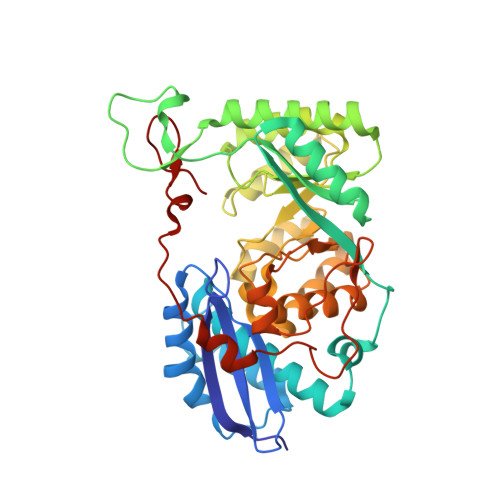 | |
UniProt | |||||
Entity Groups | |||||
| Sequence Clusters | 30% Identity50% Identity70% Identity90% Identity95% Identity100% Identity | ||||
| UniProt Group | A5KUH4 | ||||
Sequence AnnotationsExpand | |||||
Reference Sequence | |||||
| Ligands 3 Unique | |||||
|---|---|---|---|---|---|
| ID | Chains | Name / Formula / InChI Key | 2D Diagram | 3D Interactions | |
| EPE Download:Ideal Coordinates CCD File | F [auth A], L [auth C] | 4-(2-HYDROXYETHYL)-1-PIPERAZINE ETHANESULFONIC ACID C8 H18 N2 O4 S JKMHFZQWWAIEOD-UHFFFAOYSA-N |  | ||
| D8T Download:Ideal Coordinates CCD File | G [auth A], J [auth B], M [auth C], O [auth D] | D-arabinonic acid C5 H10 O6 QXKAIJAYHKCRRA-JJYYJPOSSA-N |  | ||
| MG Download:Ideal Coordinates CCD File | E [auth A], H [auth B], I [auth B], K [auth C], N [auth D] | MAGNESIUM ION Mg JLVVSXFLKOJNIY-UHFFFAOYSA-N |  | ||
| Length ( Å ) | Angle ( ˚ ) |
|---|---|
| a = 163.372 | α = 90 |
| b = 163.372 | β = 90 |
| c = 110.412 | γ = 90 |
| Software Name | Purpose |
|---|---|
| ADSC | data collection |
| BALBES | phasing |
| PHENIX | refinement |
| DENZO | data reduction |
| SCALEPACK | data scaling |


