3S8Q

Crystal structure of the R-M controller protein C.Esp1396I OL operator complex


Experimental Data Snapshot

  • Method: X-RAY DIFFRACTION
  • Resolution: 2.10 Å
  • R-Value Free: 0.219 
  • R-Value Work: 0.157 
  • R-Value Observed: 0.160 

wwPDB Validation   3D Report Full Report


This is version 1.2 of the entry. See complete history


Literature

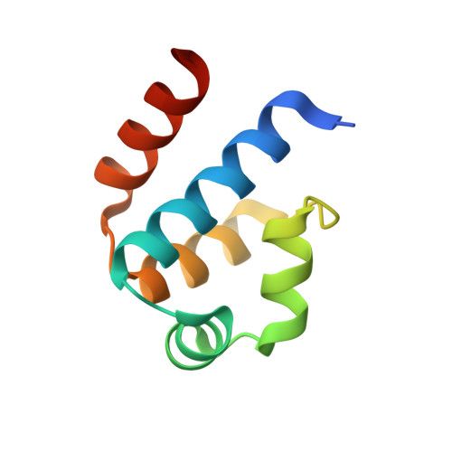
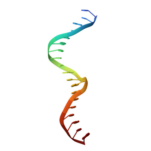
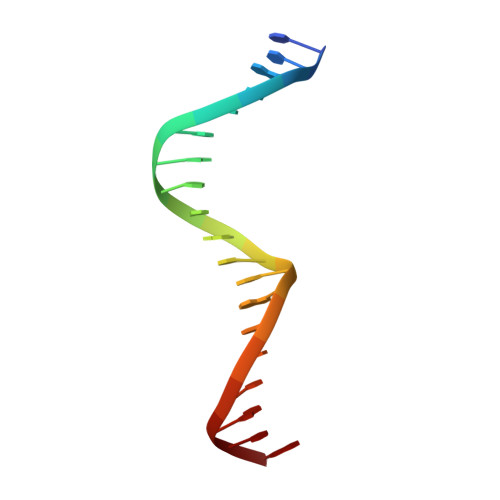
Recognition of dual symmetry by the controller protein C.Esp1396I based on the structure of the transcriptional activation complex.

McGeehan, J.E.Ball, N.J.Streeter, S.D.Thresh, S.J.Kneale, G.G.

(2012) Nucleic Acids Res 40: 4158-4167

  • DOI: https://doi.org/10.1093/nar/gkr1250
  • Primary Citation of Related Structures:  
    3S8Q

  • PubMed Abstract: 

    The controller protein C.Esp1396I regulates the timing of gene expression of the restriction-modification (RM) genes of the RM system Esp1396I. The molecular recognition of promoter sequences by such transcriptional regulators is poorly understood, in part because the DNA sequence motifs do not conform to a well-defined symmetry. We report here the crystal structure of the controller protein bound to a DNA operator site. The structure reveals how two different symmetries within the operator are simultaneously recognized by the homo-dimeric protein, underpinned by a conformational change in one of the protein subunits. The recognition of two different DNA symmetries through movement of a flexible loop in one of the protein subunits may represent a general mechanism for the recognition of pseudo-symmetric DNA sequences.


  • Organizational Affiliation

    Biomolecular Structure Group, Institute of Biomedical and Biomolecular Sciences, School of Biological Sciences, University of Portsmouth, Portsmouth, Hampshire PO1 2DY, UK.


Macromolecules

Find similar proteins by:  (by identity cutoff)  |  3D Structure
Entity ID: 1
MoleculeChains Sequence LengthOrganismDetailsImage
R-M CONTROLLER PROTEIN
A, B
82Enterobacter sp. RFL1396Mutation(s): 0 
Gene Names: esp1396IC
UniProt
Find proteins for Q8GGH0 (Enterobacter sp. RFL1396)
Explore Q8GGH0 
Go to UniProtKB:  Q8GGH0
Entity Groups  
Sequence Clusters30% Identity50% Identity70% Identity90% Identity95% Identity100% Identity
UniProt GroupQ8GGH0
Sequence Annotations
Expand
  • Reference Sequence

Find similar nucleic acids by:  Sequence   |   3D Structure  

Entity ID: 2
MoleculeChains LengthOrganismImage
DNA (5'-D(*AP*TP*GP*TP*GP*AP*CP*TP*TP*AP*TP*AP*GP*TP*CP*CP*GP*TP*G)-3')19N/A
Sequence Annotations
Expand
  • Reference Sequence

Find similar nucleic acids by:  Sequence   |   3D Structure  

Entity ID: 3
MoleculeChains LengthOrganismImage
DNA (5'-D(*TP*CP*AP*CP*GP*GP*AP*CP*TP*AP*TP*AP*AP*GP*TP*CP*AP*CP*A)-3')19N/A
Sequence Annotations
Expand
  • Reference Sequence
Experimental Data & Validation

Experimental Data

  • Method: X-RAY DIFFRACTION
  • Resolution: 2.10 Å
  • R-Value Free: 0.219 
  • R-Value Work: 0.157 
  • R-Value Observed: 0.160 
  • Space Group: P 2 21 21
Unit Cell:
Length ( Å )Angle ( ˚ )
a = 44.316α = 90
b = 61.546β = 90
c = 113.687γ = 90
Software Package:
Software NamePurpose
XSCALEdata scaling
PHASERphasing
REFMACrefinement
PDB_EXTRACTdata extraction
MxCuBEdata collection
XDSdata reduction

Structure Validation

View Full Validation Report


Entry History 

Deposition Data

Revision History  (Full details and data files)

  • Version 1.0: 2012-01-18
    Type: Initial release
  • Version 1.1: 2012-07-25
    Changes: Database references
  • Version 1.2: 2024-02-28
    Changes: Data collection, Database references