3S8F

1.8 A structure of ba3 cytochrome c oxidase from Thermus thermophilus in lipid environment


Experimental Data Snapshot

  • Method: X-RAY DIFFRACTION
  • Resolution: 1.80 Å
  • R-Value Free: 0.215 
  • R-Value Work: 0.184 
  • R-Value Observed: 0.186 

wwPDB Validation   3D Report Full Report


Ligand Structure Quality Assessment 


This is version 1.3 of the entry. See complete history


Literature

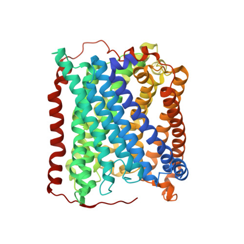
High resolution structure of the ba3 cytochrome c oxidase from Thermus thermophilus in a lipidic environment.

Tiefenbrunn, T.Liu, W.Chen, Y.Katritch, V.Stout, C.D.Fee, J.A.Cherezov, V.

(2011) PLoS One 6: e22348-e22348

  • DOI: https://doi.org/10.1371/journal.pone.0022348
  • Primary Citation of Related Structures:  
    3S8F, 3S8G

  • PubMed Abstract: 

    The fundamental chemistry underpinning aerobic life on Earth involves reduction of dioxygen to water with concomitant proton translocation. This process is catalyzed by members of the heme-copper oxidase (HCO) superfamily. Despite the availability of crystal structures for all types of HCO, the mode of action for this enzyme is not understood at the atomic level, namely how vectorial H(+) and e(-) transport are coupled. Toward addressing this problem, we report wild type and A120F mutant structures of the ba(3)-type cytochrome c oxidase from Thermus thermophilus at 1.8 Å resolution. The enzyme has been crystallized from the lipidic cubic phase, which mimics the biological membrane environment. The structures reveal 20 ordered lipid molecules that occupy binding sites on the protein surface or mediate crystal packing interfaces. The interior of the protein encloses 53 water molecules, including 3 trapped in the designated K-path of proton transfer and 8 in a cluster seen also in A-type enzymes that likely functions in egress of product water and proton translocation. The hydrophobic O(2)-uptake channel, connecting the active site to the lipid bilayer, contains a single water molecule nearest the Cu(B) atom but otherwise exhibits no residual electron density. The active site contains strong electron density for a pair of bonded atoms bridging the heme Fe(a3) and Cu(B) atoms that is best modeled as peroxide. The structure of ba(3)-oxidase reveals new information about the positioning of the enzyme within the membrane and the nature of its interactions with lipid molecules. The atomic resolution details provide insight into the mechanisms of electron transfer, oxygen diffusion into the active site, reduction of oxygen to water, and pumping of protons across the membrane. The development of a robust system for production of ba(3)-oxidase crystals diffracting to high resolution, together with an established expression system for generating mutants, opens the door for systematic structure-function studies.


  • Organizational Affiliation

    Department of Molecular Biology, The Scripps Research Institute, La Jolla, California, United States of America.


Macromolecules
Find similar proteins by:  (by identity cutoff)  |  3D Structure
Entity ID: 1
MoleculeChains Sequence LengthOrganismDetailsImage
Cytochrome c oxidase subunit 1569Thermus thermophilus HB8Mutation(s): 0 
Gene Names: cbaATTHA1135
EC: 1.9.3.1
Membrane Entity: Yes 
UniProt
Find proteins for Q5SJ79 (Thermus thermophilus (strain ATCC 27634 / DSM 579 / HB8))
Explore Q5SJ79 
Go to UniProtKB:  Q5SJ79
Entity Groups  
Sequence Clusters30% Identity50% Identity70% Identity90% Identity95% Identity100% Identity
UniProt GroupQ5SJ79
Sequence Annotations
Expand
  • Reference Sequence
Find similar proteins by:  (by identity cutoff)  |  3D Structure
Entity ID: 2
MoleculeChains Sequence LengthOrganismDetailsImage
Cytochrome c oxidase subunit 2168Thermus thermophilus HB8Mutation(s): 0 
Gene Names: cbaBcbacctaCTTHA1134
EC: 1.9.3.1
Membrane Entity: Yes 
UniProt
Find proteins for Q5SJ80 (Thermus thermophilus (strain ATCC 27634 / DSM 579 / HB8))
Explore Q5SJ80 
Go to UniProtKB:  Q5SJ80
Entity Groups  
Sequence Clusters30% Identity50% Identity70% Identity90% Identity95% Identity100% Identity
UniProt GroupQ5SJ80
Sequence Annotations
Expand
  • Reference Sequence
Find similar proteins by:  (by identity cutoff)  |  3D Structure
Entity ID: 3
MoleculeChains Sequence LengthOrganismDetailsImage
Cytochrome c oxidase polypeptide 2A34Thermus thermophilus HB8Mutation(s): 0 
Gene Names: cbaDTTHA1133
EC: 1.9.3.1
Membrane Entity: Yes 
UniProt
Find proteins for P82543 (Thermus thermophilus (strain ATCC 27634 / DSM 579 / HB8))
Explore P82543 
Go to UniProtKB:  P82543
Entity Groups  
Sequence Clusters30% Identity50% Identity70% Identity90% Identity95% Identity100% Identity
UniProt GroupP82543
Sequence Annotations
Expand
  • Reference Sequence
Small Molecules
Ligands 6 Unique
IDChains Name / Formula / InChI Key2D Diagram3D Interactions
HAS
Query on HAS

Download Ideal Coordinates CCD File 
F [auth A]HEME-AS
C54 H64 Fe N4 O6
PDYODZVCODUKFH-ZOMLSHGTSA-L
HEM
Query on HEM

Download Ideal Coordinates CCD File 
E [auth A]PROTOPORPHYRIN IX CONTAINING FE
C34 H32 Fe N4 O4
KABFMIBPWCXCRK-RGGAHWMASA-L
OLC
Query on OLC

Download Ideal Coordinates CCD File 
H [auth A]
I [auth A]
J [auth A]
K [auth A]
L [auth A]
H [auth A],
I [auth A],
J [auth A],
K [auth A],
L [auth A],
M [auth A],
N [auth A],
O [auth A],
P [auth A],
Q [auth A],
R [auth A],
T [auth B],
U [auth B],
V [auth B],
W [auth B],
X [auth C]
(2R)-2,3-dihydroxypropyl (9Z)-octadec-9-enoate
C21 H40 O4
RZRNAYUHWVFMIP-GDCKJWNLSA-N
CUA
Query on CUA

Download Ideal Coordinates CCD File 
S [auth B]DINUCLEAR COPPER ION
Cu2
ALKZAGKDWUSJED-UHFFFAOYSA-N
CU
Query on CU

Download Ideal Coordinates CCD File 
D [auth A]COPPER (II) ION
Cu
JPVYNHNXODAKFH-UHFFFAOYSA-N
PER
Query on PER

Download Ideal Coordinates CCD File 
G [auth A]PEROXIDE ION
O2
ANAIPYUSIMHBEL-UHFFFAOYSA-N
Experimental Data & Validation

Experimental Data

  • Method: X-RAY DIFFRACTION
  • Resolution: 1.80 Å
  • R-Value Free: 0.215 
  • R-Value Work: 0.184 
  • R-Value Observed: 0.186 
  • Space Group: C 1 2 1
Unit Cell:
Length ( Å )Angle ( ˚ )
a = 143.585α = 90
b = 97.816β = 128.3
c = 94.952γ = 90
Software Package:
Software NamePurpose
DENZOdata reduction
SCALEPACKdata scaling
REFMACrefinement
PDB_EXTRACTdata extraction
HKL-2000data collection
HKL-2000data reduction
HKL-2000data scaling
MiFitphasing

Structure Validation

View Full Validation Report


Ligand Structure Quality Assessment 


Entry History 

Deposition Data

Revision History  (Full details and data files)

  • Version 1.0: 2011-08-03
    Type: Initial release
  • Version 1.1: 2012-03-21
    Changes: Database references
  • Version 1.2: 2017-11-08
    Changes: Refinement description
  • Version 1.3: 2024-02-28
    Changes: Data collection, Database references, Derived calculations